Revision Notes
OCR A Level Biology: Exchange and Transport (H420) – Revision Notes
Condensed recall notes on exchange surfaces, transport in animals and transport in plants for OCR A Level Biology A (H420), Module 3.
- Subject
- Biology
- Level
- A LEVELS
- Topic
- Exchange and transport
- Author
- Marlbridge Academic Team
- Updated
- Reviewed by
- Hina Mogul (what this means)
Aligned to OCR A Level Biology (H420), Version 4.1 (April 2026), for first assessment in 2025. Official specification .
Syllabus page (what it covers and how it is assessed): OCR A Level Biology.
Syllabus points this page covers
H420
- 3 Exchange and transport (whole topic)
- 3.1.1 Exchange surfaces
- 3.1.2 Transport in animals
- 3.1.3 Transport in plants
Found an error? Report a correction.
Need help with this topic? Request a free trial class for A Level Biology (H420).
These notes condense Module 3 (sections 3.1.1 to 3.1.3) of the OCR A Level Biology A (H420) specification, Version 4.1 (April 2026). Use the study guide for full explanations, then test yourself with the practice questions. The course hub and printable checklist show where this fits, and the free diagnostics help you spot weak topics. Each outcome is covered, but the practical outcomes (dissection, drawing, slides) are the briefest.
3.1.1 Exchange surfaces
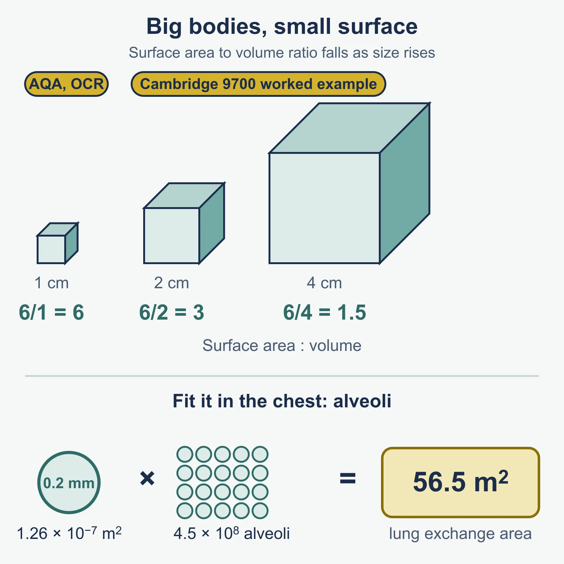
Why they are needed. SA:V = surface area ÷ volume. It falls as size rises, while metabolic activity raises demand. Single-celled organisms can use the body surface; larger or more active animals need a specialised surface plus ventilation.
Efficient exchange surface:
| Feature | Example |
|---|---|
| Large surface area | Root hair cells; folded lamellae |
| Thin layer, short distance | Alveolar wall |
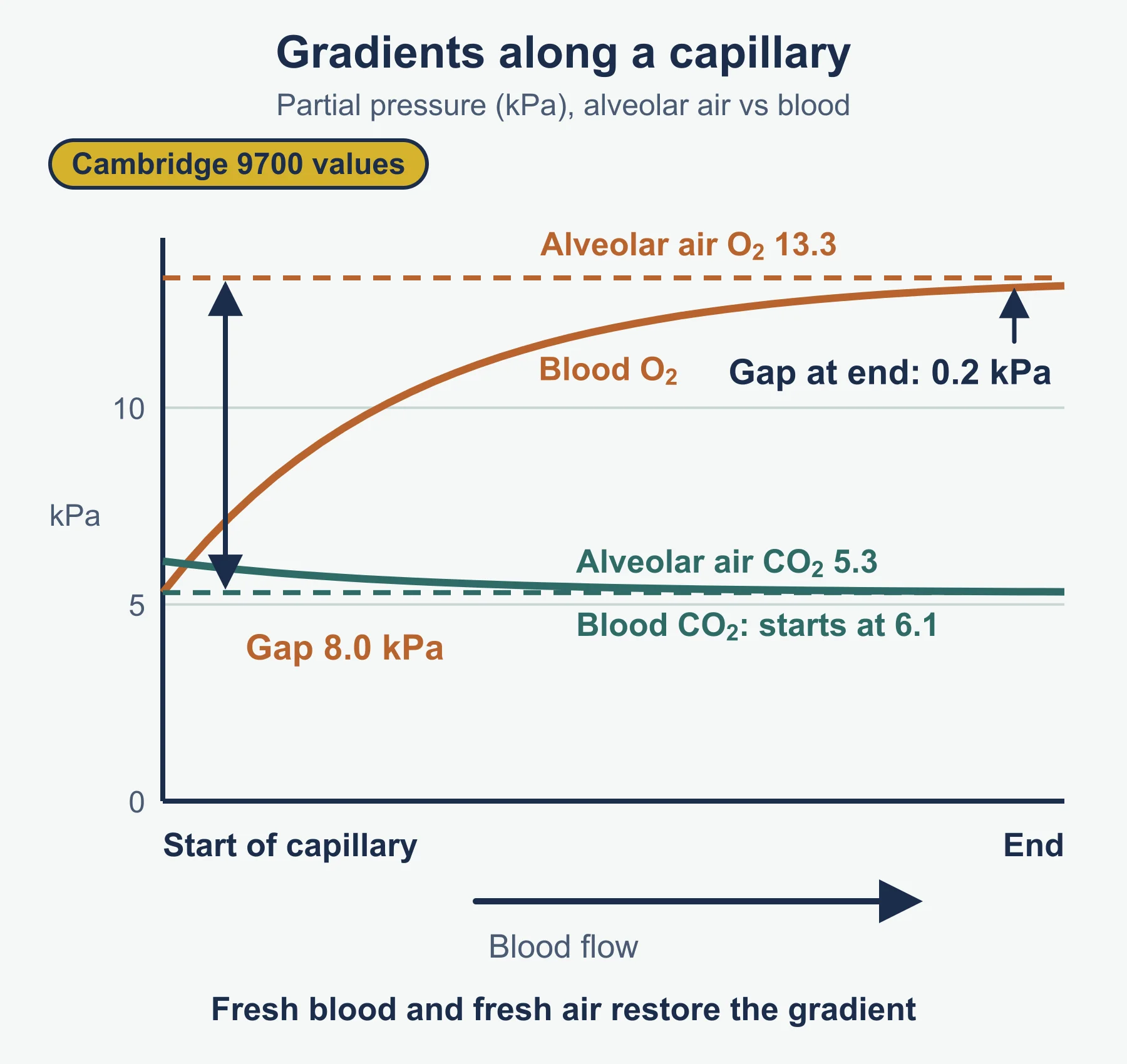
| Maintained gradient (blood flow, ventilation) | Gills, alveoli |
Mammalian system, by region:
| Tissue | Where | Function |
|---|---|---|
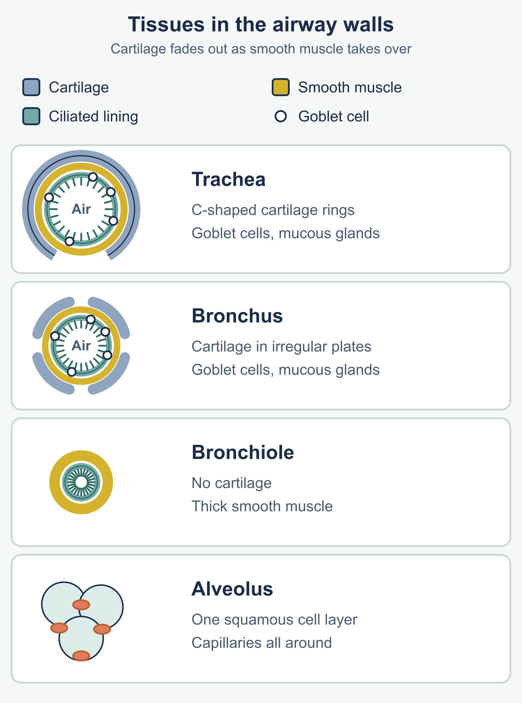
| Cartilage | Trachea (C-shaped rings), bronchi | Keeps airway open |
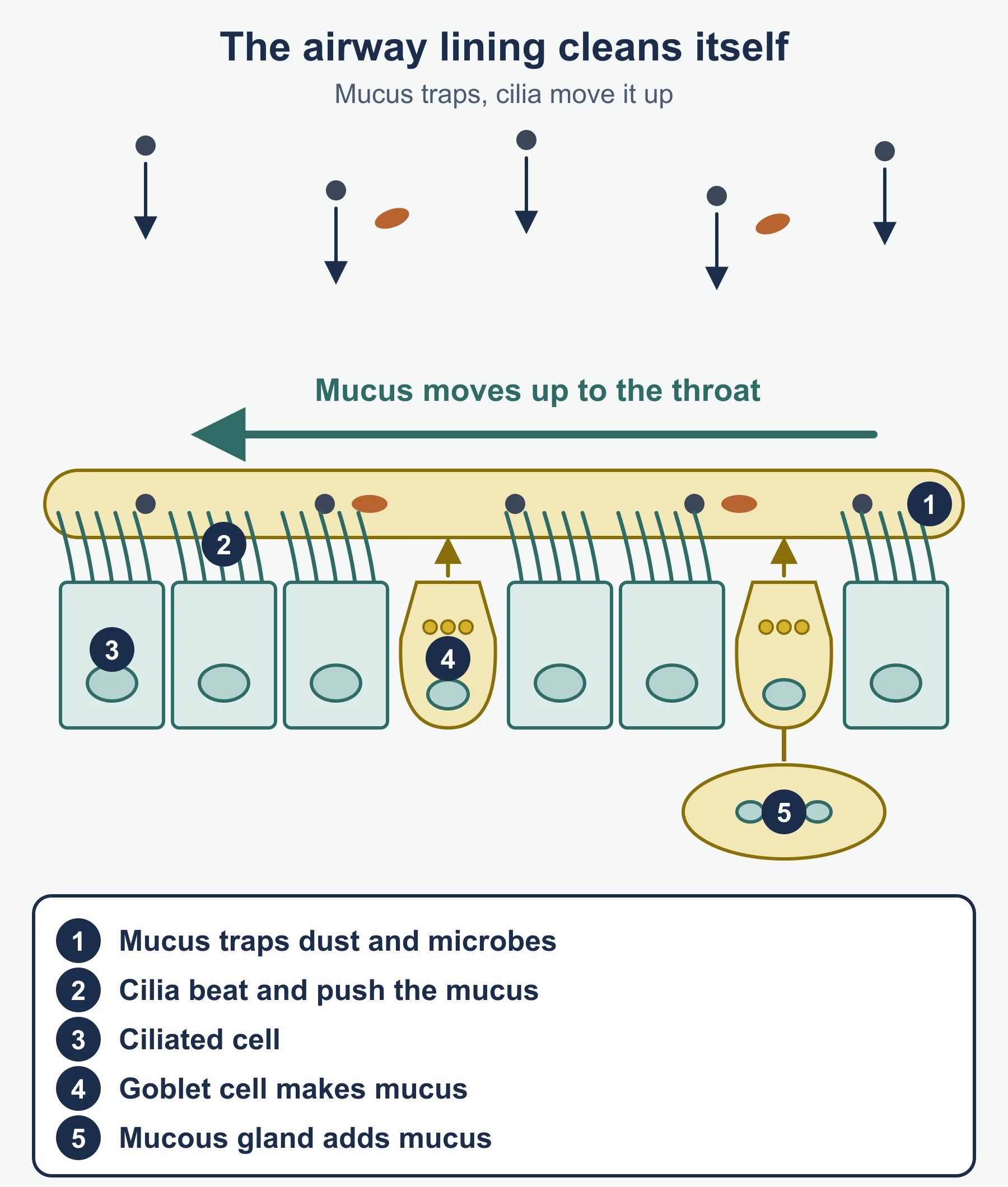
| Ciliated epithelium | Trachea to bronchioles | Sweeps mucus upward |
| Goblet cells | Trachea, bronchi | Make mucus to trap particles |
| Smooth muscle | Trachea to bronchioles | Alters airway diameter |
| Elastic fibres | All, including alveoli | Recoil, helping expiration |
The alveolus itself has no cartilage, cilia, goblet cells or smooth muscle: its wall is a thin squamous layer with elastic fibres.
Ventilation (mammal).
| Stage | Muscles | Ribs and diaphragm | Volume and pressure |
|---|---|---|---|
| Inspiration | External intercostals and diaphragm contract | Ribs up and out; diaphragm flattens | Volume up, pressure down |
| Expiration | External intercostals and diaphragm relax; internal intercostals can contract in forced breathing | Ribs down and in; diaphragm domes | Volume down, pressure up |
Lung volumes. Tidal volume is the volume per resting breath. Vital capacity is the maximum volume that can be exhaled after a maximal inhalation. Tidal volume is only part of vital capacity, so exercise can raise both tidal volume and breathing rate. Ventilation rate = tidal volume × breathing rate. A spirometer trace gives oxygen uptake from the fall in the trace over time, once carbon dioxide is removed.
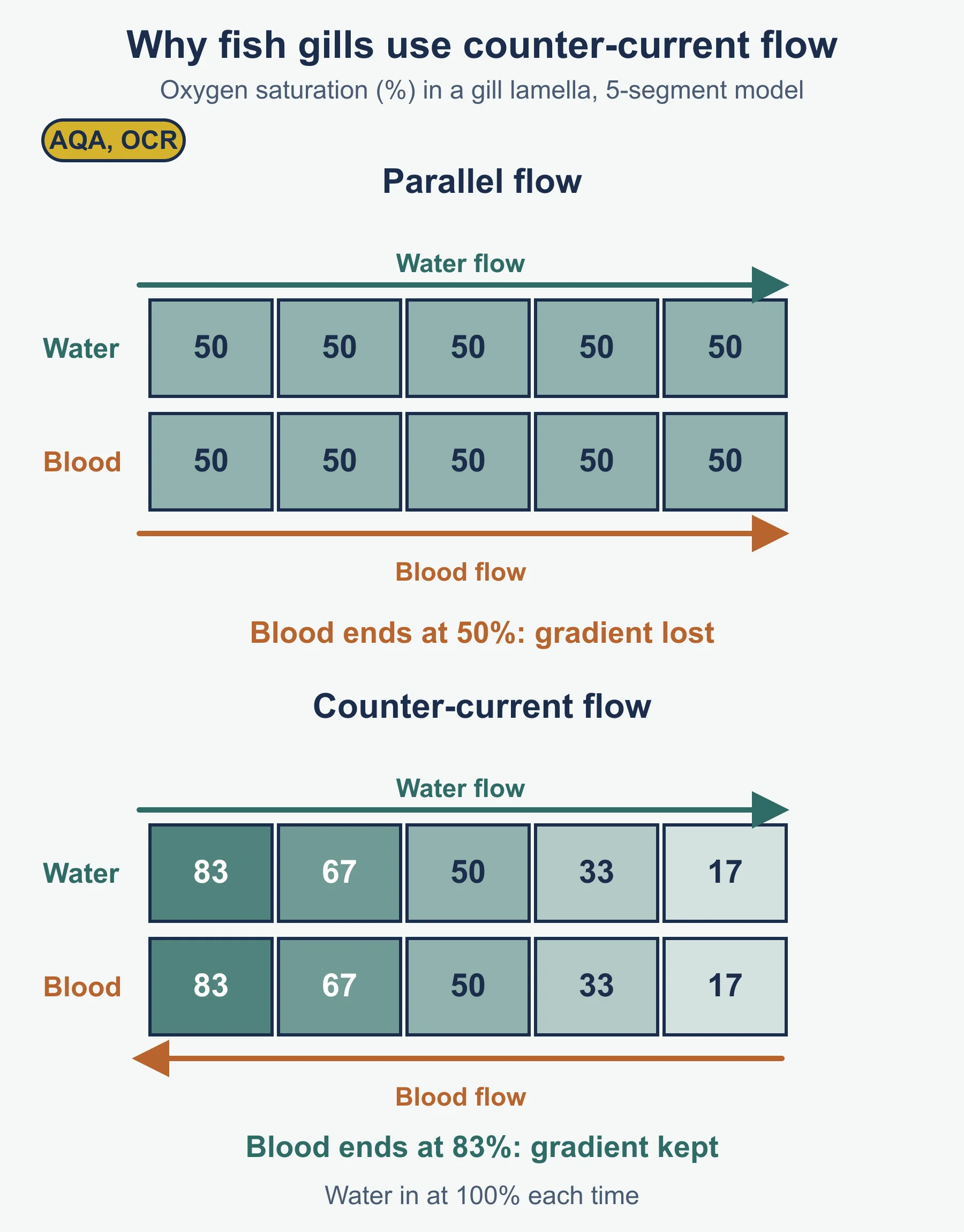
Bony fish. Lowering the buccal cavity floor raises volume, so water enters. Raising it forces water over the gills as the operculum opens. Each filament carries lamellae. Blood flows opposite to water (countercurrent), so the gradient is kept across the full length.
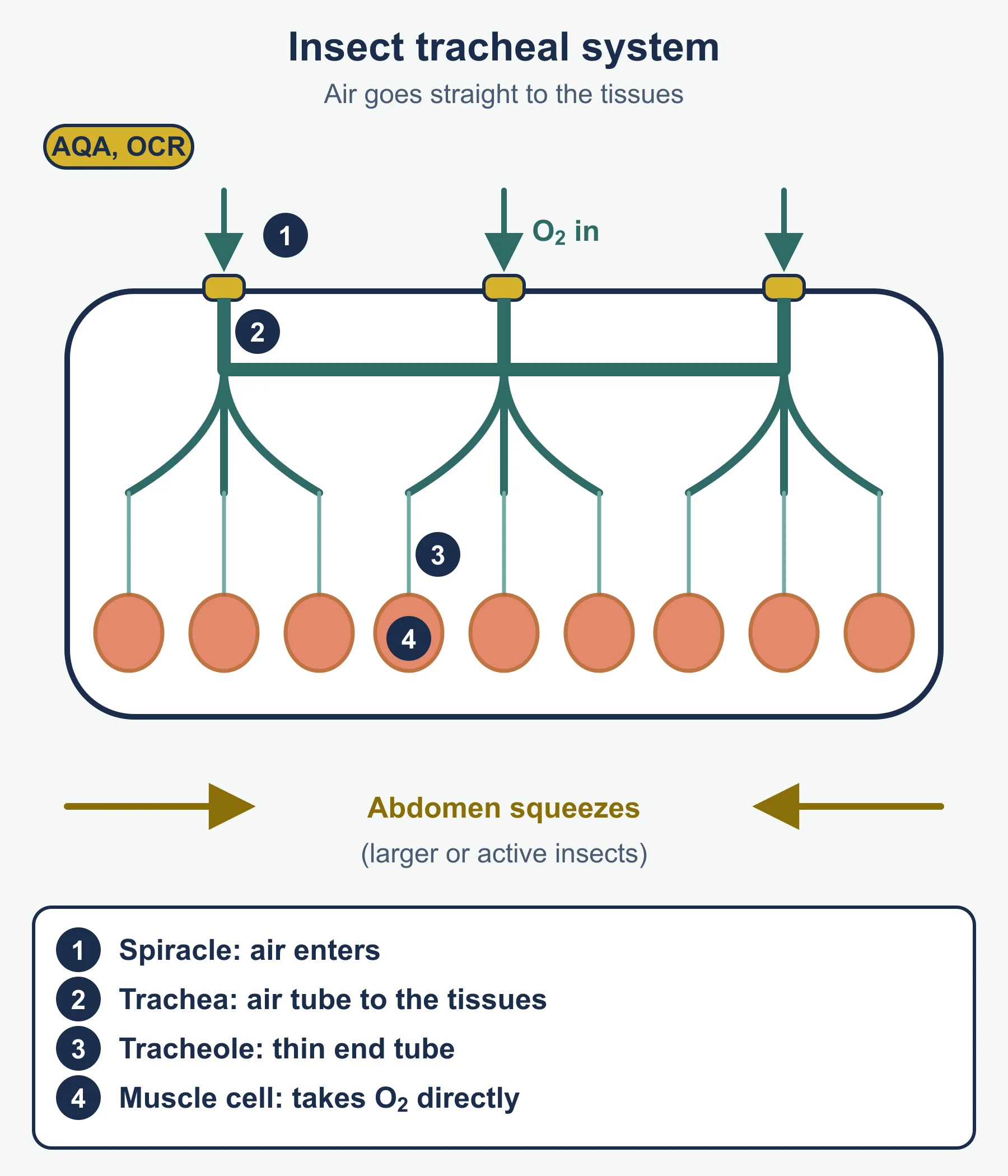
Insects. Air enters through spiracles into tracheae and tracheoles. Gas exchange takes place at tracheal fluid at the tracheole ends. Thoracic and abdominal movements change body volume to ventilate.
Practical outcomes (briefest). Fish gill or insect trachea dissection and drawing; stained microscope slides of exchange surfaces: draw only what you see, with clean lines and no shading.
3.1.2 Transport in animals
Need. Greater size, higher metabolic rate and low SA:V mean diffusion is too slow.
Circulatory systems.
| Animal | Type | Key point |
|---|---|---|
| Insect | Open | Fluid bathes tissues; low pressure |
| Fish | Closed, single | Blood passes the heart once per circuit |
| Mammal | Closed, double | Heart pumps twice per circuit, so pressure is restored after the lungs |
Vessels.
| Vessel | Wall tissues | Notes |
|---|---|---|
| Artery | Thick; elastic fibres, smooth muscle, collagen | Withstands high pressure |
| Arteriole | More smooth muscle | Controls flow |
| Capillary | Endothelium only | Short distance |
| Venule, vein | Thin wall; valves in veins | Return blood at low pressure |
Tissue fluid. Hydrostatic pressure forces fluid out at the arteriole end. Oncotic pressure from plasma proteins pulls water back. At the venule end the hydrostatic pressure has fallen, so net movement is inwards. Tissue fluid lacks most large proteins and red cells. Lymph is similar but has more lymphocytes and fewer nutrients, and is returned to the blood by lymph vessels.
Heart. External structure: atria, ventricles, coronary arteries. Internal: septum, atrioventricular valves, semilunar valves, thicker left ventricle wall. Dissection: record sizes and cut the wall to reveal the valves.
Cardiac cycle.
| Phase | Valves | Pressure |
|---|---|---|
| Atrial systole | AV open | Atria above ventricles |
| Ventricular systole | AV shut, semilunar open | Ventricle above aorta |
| Diastole | Semilunar shut, AV open | Atria fill |
Cardiac output = heart rate × stroke volume.
Coordination. SAN starts excitation across atria, AVN delays it, purkyne tissue carries it through the ventricles. Cardiac muscle is myogenic.
ECG. Normal traces are regular. Tachycardia is faster; bradycardia slower. Fibrillation is irregular. An ectopic beat comes early. Rate = 60 ÷ time between R waves.
Gas transport. Haemoglobin binds oxygen reversibly. Carbon dioxide forms carbonic acid by carbonic anhydrase, then hydrogencarbonate and H⁺. Haemoglobin binds H⁺ (haemoglobinic acid). Hydrogencarbonate leaves and chloride enters (chloride shift).
Dissociation curves. Fetal haemoglobin has a higher affinity and its curve lies to the left of adult. More carbon dioxide shifts the curve right, the Bohr effect.
3.1.3 Transport in plants
Need. Large and complex plants have low SA:V and cells far from sources.
Vascular tissue (herbaceous dicot).
| Tissue | Cells | Key features |
|---|---|---|
| Xylem | Vessels | Dead, lignified, no end walls |
| Phloem | Sieve tube elements, companion cells | Living, sieve plates; companions support |
Stem: bundles in a ring. Root: central core. Leaf: midrib bundle. Practical outcomes: stained sections and stem dissection.
Transpiration. Water vapour loss through open stomata, a consequence of gas exchange. Faster with higher temperature, light, wind and lower humidity. A potometer estimates uptake: volume = πr² × distance, divided by time.
Water movement. Root hair → cortex by apoplast (cell walls) or symplast (cytoplasm and plasmodesmata) → xylem. Evaporation from leaf cells lowers water potential; cohesion pulls the column up under tension; adhesion helps along walls; this is the transpiration stream. Water moves from higher (less negative) to lower (more negative) water potential. ψ = ψs + ψp.
Adaptations.
| Type | Example | Features |
|---|---|---|
| Xerophyte | Cactus; marram grass | Thick cuticle, rolled leaves, sunken stomata, hairs, spines, reduced leaves |
| Hydrophyte | Water lily | Air spaces, stomata on upper surface, thin cuticle |
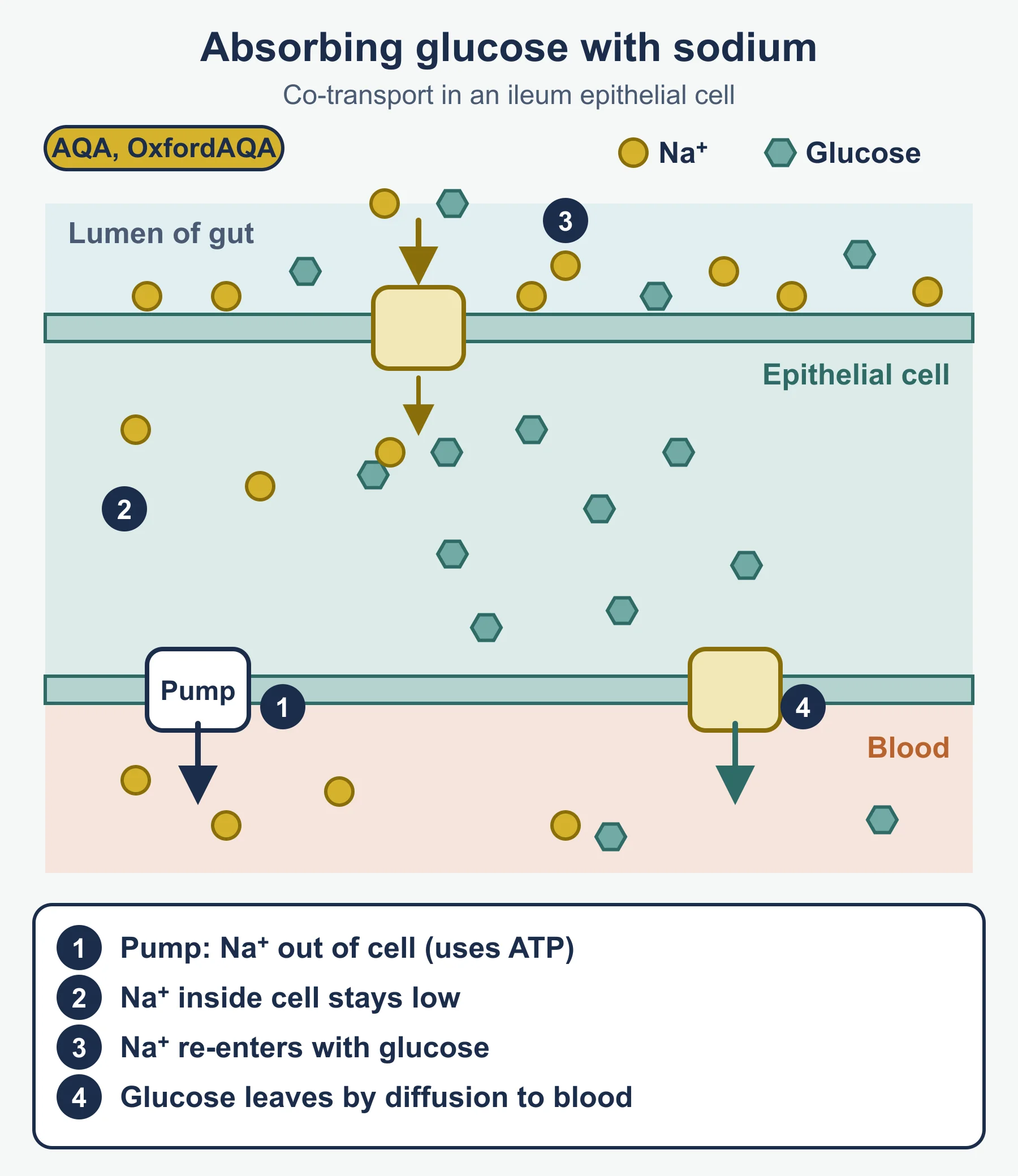
Translocation. Energy-requiring transport of sucrose from sources (leaves) to sinks (roots, meristems). Active loading: companion cells pump H⁺ out using ATP; H⁺ returns with sucrose via co-transport; sieve tube water potential falls, water enters, hydrostatic pressure rises, sap flows. At the sink, sucrose is removed and used or stored.
Methods in steps
Explaining an exchange surface. (1) Name the feature. (2) State what it does to distance, area or gradient. (3) Say how that changes the rate of diffusion. (4) Repeat for each feature the question asks about.
Reading an ECG. (1) Find the time between two successive R waves, in seconds. (2) Divide 60 by it. (3) Compare with the usual resting range of about 60 to 100 beats per minute. (4) Look at the spacing: regular, irregular, or one early beat.
Tissue fluid questions. (1) Subtract oncotic from hydrostatic pressure. (2) A positive answer means fluid leaves the capillary; a negative one means it returns. (3) Link the change in hydrostatic pressure along the capillary to the change in direction. (4) Say what happens to excess fluid: it drains into lymph vessels.
Potometer calculations. (1) Convert all lengths to the same unit. (2) Volume = πr² × distance moved. (3) Divide by the time taken. (4) State that the apparatus measures uptake, which only estimates transpiration.
Must-know distinctions
| Compare | First | Second |
|---|---|---|
| Open and closed circulation | Fluid bathes tissues directly | Blood stays in vessels |
| Single and double circulation | One pass through the heart | Two passes, so pressure is restored |
| Tissue fluid and plasma | Few large proteins | Contains plasma proteins |
| Hydrostatic and oncotic pressure | Pushes fluid out | Pulls water in |
| Apoplast and symplast | Walls and spaces | Cytoplasm through plasmodesmata |
| Xylem and phloem | Water and ions, upward, dead cells | Assimilates, either direction, living cells |
| Xerophyte and hydrophyte | Reduces loss of water | Floats and exchanges gases through upper surface |
| Source and sink | Where sucrose is loaded | Where sucrose is removed |
Countercurrent reminder
Imagine water that loses oxygen from 80% to 40% saturation along a lamella. Blood moving the other way enters at 30% and leaves at 70%. At every point along the lamella, water carries about 10 percentage points more oxygen than the blood beside it, so diffusion carries on throughout. With parallel flow, the two would meet at an intermediate saturation, about 55%, and net uptake would stop partway along.
Quick self-test
- A cube has sides of 0.5 mm. Calculate its SA:V, then for 5 mm.
- Heart rate 72, stroke volume 70 cm³: find cardiac output.
- R waves are 1.2 s apart: find the rate and name it.
- Tidal volume 0.5 dm³, 14 breaths per minute: find ventilation rate.
- Hydrostatic pressure is 4.0 kPa, oncotic 3.0 kPa: state the net pressure out.
- A cell has ψs of −900 kPa and ψp of +400 kPa: find ψ.
- Name the cells that transport sucrose.
- Which way does more carbon dioxide shift the curve?
- Name the enzyme in red cells that forms carbonic acid.
- A bubble moves 36 mm in 5 minutes in a tube of radius 0.5 mm: find uptake per minute.
Answers
- 12 mm⁻¹ and 1.2 mm⁻¹.
- 72 × 70 = 5040 cm³ per minute.
- 60 ÷ 1.2 = 50 beats per minute: bradycardia.
- 7.0 dm³ per minute.
- 1.0 kPa.
- −500 kPa.
- Sieve tube elements, helped by companion cells.
- Right.
- Carbonic anhydrase.
- π × 0.5² × 36 = 28.3 mm³, so 5.65 mm³ per minute.
Where marks are usually lost
- Naming a feature without explaining the effect on diffusion.
- Mixing up the external and internal intercostal muscles.
- Saying “air is sucked in” instead of describing pressure falling.
- Forgetting that fish blood flows opposite to water.
- Not stating the direction of net movement in tissue fluid.
- Mixing up which cardiac valves are shut in each phase.
- Giving heart rate without using the time between R waves.
- Forgetting that xylem is dead and phloem is living.
- Quoting water movement from “high to low” without the sign.
Frequently asked questions
What do the external and internal intercostal muscles do? During normal inspiration the external intercostal muscles contract to raise the ribcage, while the internal intercostal muscles are used in forced expiration. Mixing up the two is a frequent error in gas exchange answers.
How should inspiration be described without saying air is “sucked in”? Describe the volume of the thorax increasing, so the pressure inside falls below atmospheric pressure, and air then moves into the lungs down the pressure gradient.
What is the countercurrent system in fish gills? In fish gills, blood flows in the opposite direction to the water passing over the lamellae. This keeps a diffusion gradient along the whole length of the lamella, so more oxygen is taken up than with parallel flow.
How do you calculate heart rate from an ECG? Measure the time between two R waves, which is the time for one heartbeat, and divide 60 seconds by it to give beats per minute.
What is the main difference between xylem and phloem? Xylem vessels are dead tissue that carry water and mineral ions upwards from the roots, while phloem sieve tubes are living tissue that transport assimilates such as sucrose in both directions. These notes cover Module 3 of OCR A Level Biology A (H420).
Official syllabus
OCR Advanced GCE in Biology A (H420), Version 4.1 (April 2026), for first assessment in 2025, published by OCR.
Get free revision emails (optional)
Occasional emails with practice questions, worked explanations and links to free resources for the qualification and subjects you choose. No spam, and you can unsubscribe from any email. The free tools on this site never need an email.
Related resources
-
Study guides
OCR A Level Biology: Exchange and Transport (H420)
OCR A Level Biology A (H420) Module 3 guide to exchange surfaces, gas exchange, the heart, haemoglobin and transport in plants, with common mistakes to avoid.
Biology · OCR · A Level
Read guide -
Practice questions
OCR A Level Biology: Exchange and Transport (H420) – Practice Questions
Twelve original worked questions on gas exchange, the heart, haemoglobin and plant transport for OCR A Level Biology A (H420), Module 3.
Biology · OCR · A Level
Try questions -
Study guides
OCR A Level Biology: Foundations in Biology (H420)
Cell structure, biological molecules, and nucleotides and nucleic acids – the opening content of Module 2 for OCR A Level Biology A (H420), Version 4.1.
Biology · OCR · A Level
Read guide
Related articles
-
study skills
How to revise for a science examination
Most science revision fails because it rereads notes instead of retrieving them. A practical method for revising physics, chemistry and biology in the weeks before a paper.
14 July 2026
-
teaching
A Level tuition in Dubai: Cambridge or Pearson Edexcel International A Level?
Schools in Dubai and Abu Dhabi teach either Cambridge International AS & A Level or Pearson Edexcel International A Level. How the two are structured, why it matters for tuition, and how online classes from Lahore work.
9 October 2026
Studying this with a teacher
Working through Biology A LEVELS?
This page is free and stays free. If you would rather be taught it, Marlbridge runs Biology classes one-to-one, online in your own time zone. One-to-one only for this course: Rs 6,000 per class in Pakistan, with other currencies on the fees page. The first trial class is free. WhatsApp replies within an hour (8am–11pm Pakistan time, every day); email the same day.
OCR Biology teachers at Marlbridge