
Revision Notes
AQA A-level Biology: Cell Recognition and the Immune System (7402) – Revision Notes
Condensed AQA 7402 notes on 3.2.4: antigens, phagocytosis, T and B cells, antibodies, vaccines, HIV, monoclonal antibodies and ELISA, for A-level Biology.
- Subject
- Biology
- Level
- A LEVEL (AS CONTENT)
- Topic
- Cell Recognition and the Immune System
- Author
- Marlbridge Academic Team
- Updated
- Reviewed by
- Hina Mogul (what this means)
Aligned to AQA A Level Biology (7402), For first teaching 2015. Official specification .
Syllabus page (what it covers and how it is assessed): AQA A Level Biology.
Syllabus points this page covers
7402 (AS content of the A Level)
- 3.2.4 Cell recognition and the immune system
This page covers 1 of the 6 subtopics recorded here in topic 2, Cells.
Found an error? Report a correction.
Need help with this topic? Request a free trial class for A Level Biology (7402).
Short notes for the final weeks. These notes follow section 3.2.4, Cell recognition and the immune system, in the AQA AS and A-level Biology specification (A-level 7402), for first teaching 2015, exams June 2016 onwards. The section is part of the AS content, so nothing here is marked A-level only. Find the AQA A-level Biology hub, tick off topics on the checklist, and use the free diagnostics to find weak spots. Virus structure and protein structure are in the cell structure notes and the lipids and protein structure notes.
Key definitions
| Term | Meaning |
|---|---|
| Antigen | A molecule, usually a protein on a cell surface, that is recognised as foreign and triggers an immune response |
| Antibody | A protein made by B cells (secreted by plasma cells) that binds specifically to one antigen |
| Antigen-presenting cell (APC) | A cell that displays foreign antigens on its surface, for example a phagocyte after engulfing a pathogen |
| Phagocyte | A white blood cell that engulfs and digests pathogens |
| Clonal selection | Activation of the particular B cell (or T cell) whose receptors are complementary to the antigen, so that only it divides |
| Plasma cell | A cell formed from an activated B cell that secretes large amounts of one antibody |
| Memory cell | A long-lived lymphocyte that responds quickly if the same antigen appears again |
| Monoclonal antibodies | Identical antibodies made by one clone of plasma cells, all binding the same antigen |
| Herd immunity | Protection of unvaccinated people because so many others are immune that the pathogen cannot spread easily |
| Active immunity | Immunity in which the body makes its own antibodies and memory cells |
| Passive immunity | Immunity from antibodies given to the body from outside; no memory cells are made |
Cell recognition
Every cell type carries specific molecules on its surface, many of them proteins, that identify it. These let the immune system recognise:
- pathogens (bacteria, viruses, fungi);
- cells from other organisms of the same species (for example transplanted organs or transfused blood);
- abnormal body cells (for example cancer cells or virus-infected cells);
- toxins.
Antigen variability
Some pathogens change their surface antigens often, for example the influenza virus. When this happens:
- memory cells from an earlier infection or vaccine no longer have complementary receptors;
- the next infection produces a slow primary response, so people become ill again;
- a vaccine may need to be changed regularly, and one vaccine cannot cover every strain.
Phagocytosis
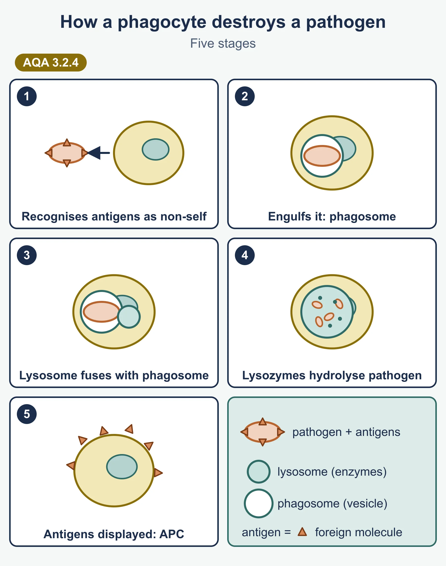
- The phagocyte recognises the foreign antigens on a pathogen and binds to it.
- It engulfs the pathogen, enclosing it in a vesicle called a phagosome.
- A lysosome fuses with the phagosome and releases hydrolytic enzymes (the specification names them lysozymes).
- The enzymes hydrolyse the pathogen.
- The phagocyte displays the pathogen’s antigens on its surface, becoming an antigen-presenting cell.
The cellular response (T lymphocytes)
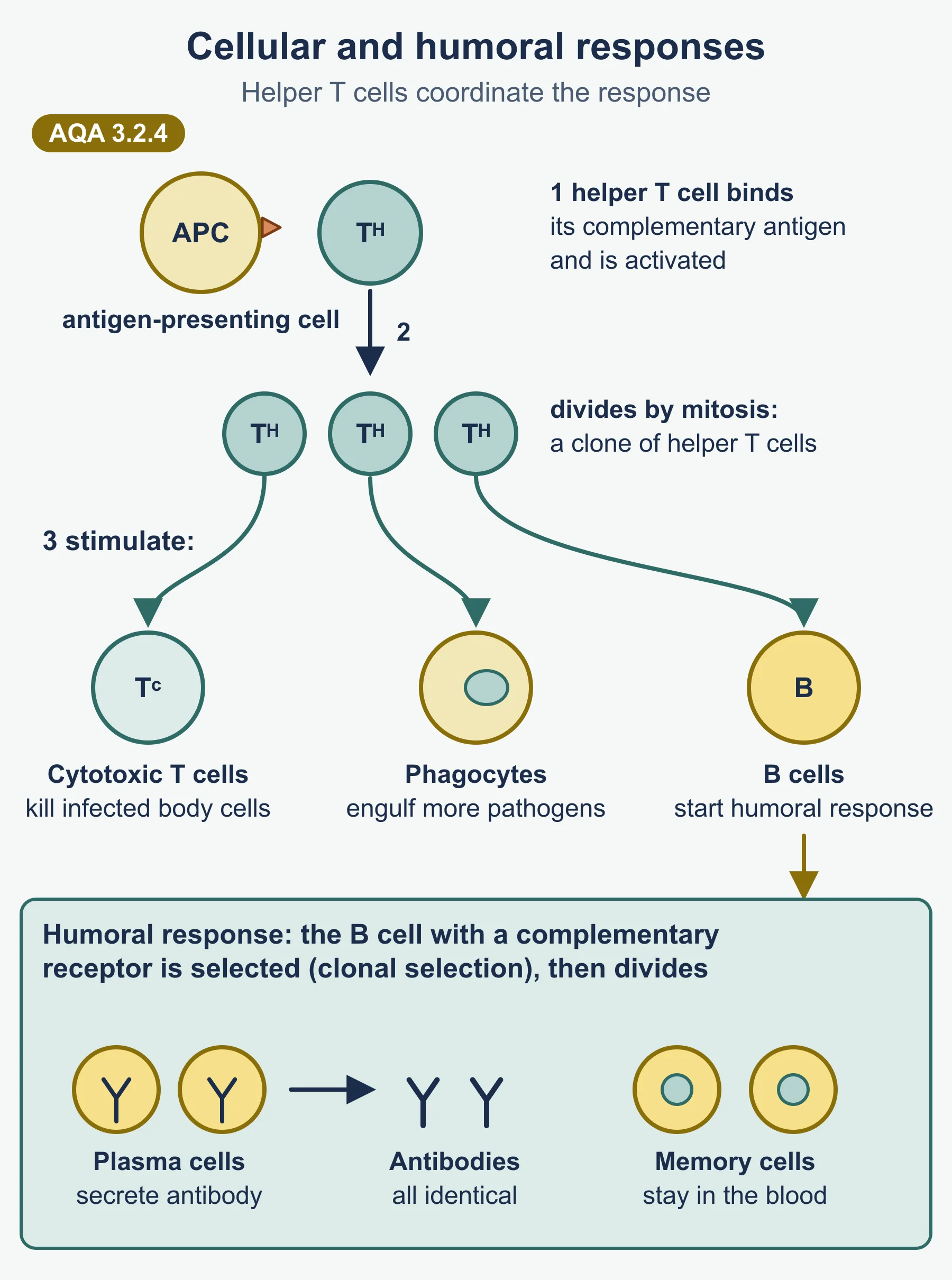
- Antigen-presenting cells (phagocytes, infected body cells, cancer cells or transplanted cells) show foreign antigens.
- A helper T cell (TH cell) with a receptor complementary to that antigen binds to it and is activated.
- The TH cell divides by mitosis to form a clone.
- TH cells then stimulate:
- cytotoxic T cells (TC cells), which kill infected body cells (for example by making holes in their membranes);
- B cells, starting the humoral response;
- phagocytes, to engulf more pathogens.
The roles of other T cells are not required.
The humoral response (B lymphocytes)
- Each B cell has antibodies on its surface, complementary to one antigen. There are millions of different B cells.
- The B cell with complementary antibodies binds the antigen, takes it in and presents it.
- A helper T cell binds to the presented antigen and activates the B cell. This is clonal selection.
- The activated B cell divides by mitosis into a clone.
- The clone differentiates into plasma cells, which secrete large amounts of one antibody (monoclonal antibodies), and memory cells.
Antibody structure
| Part | Description |
|---|---|
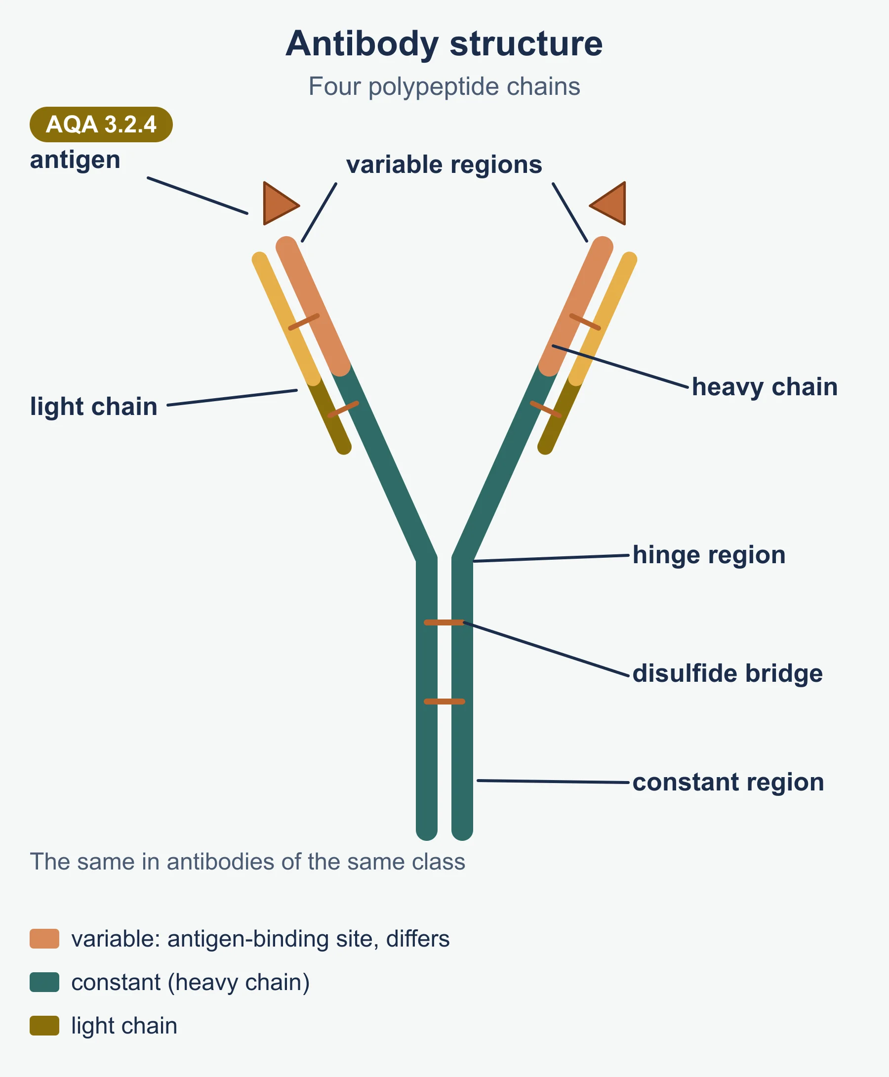
| Chains | Four polypeptide chains: two heavy and two light, held together by disulfide bridges (quaternary structure) |
| Variable region | Different in each type of antibody; its shape forms the antigen-binding site, complementary to one antigen |
| Constant region | The same in all antibodies of a class; binds to receptors on cells such as phagocytes |
| Hinge region | Lets the two arms move, so the antibody can bind two antigens a different distance apart |
Each antibody has two antigen-binding sites.
Antigen-antibody complexes
- Agglutination: each antibody can bind two bacteria at once, so bacteria clump together.
- Phagocytosis: the clumped bacteria, marked by antibodies, are engulfed and destroyed by phagocytes.
Primary and secondary responses
| Primary response | Secondary response | |
|---|---|---|
| When | First exposure to the antigen | Later exposure to the same antigen |
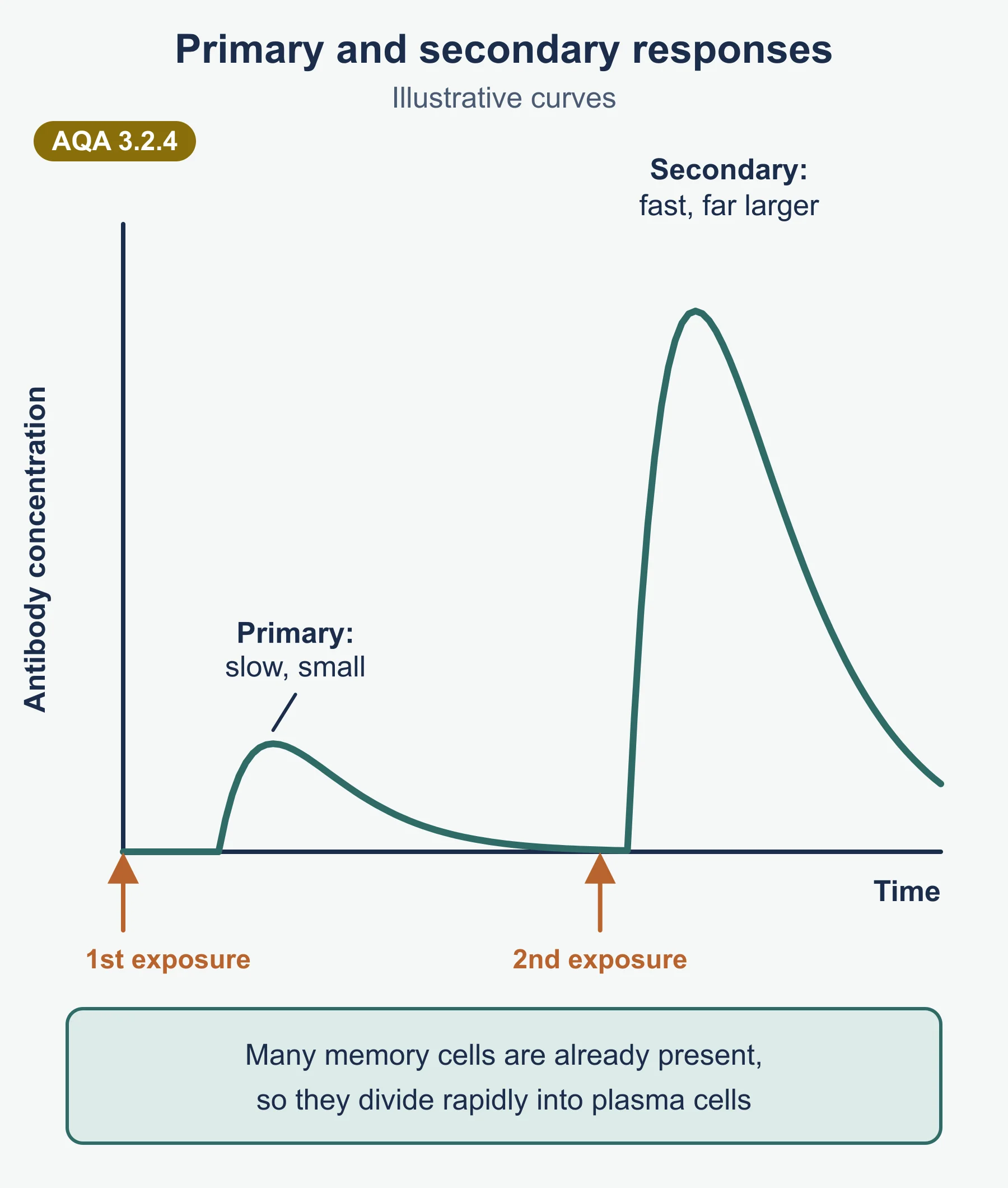
| Speed | Slow: few B and T cells have the right receptors at first | Fast: many memory cells are ready |
| Antibody level | Lower | Much higher, and reached sooner |
| Symptoms | Usually develop | Usually none; the pathogen is destroyed first |
| Cells involved | Plasma cells make antibodies; memory cells formed | Memory B cells divide rapidly into plasma cells; memory T cells into TH and TC cells |
Vaccines and herd immunity
- A vaccine contains antigens, for example in dead or weakened (attenuated) pathogens, or as isolated antigens.
- It triggers a primary response, so memory cells are made. A later infection causes a rapid secondary response.
- For individuals: protection without having the disease first.
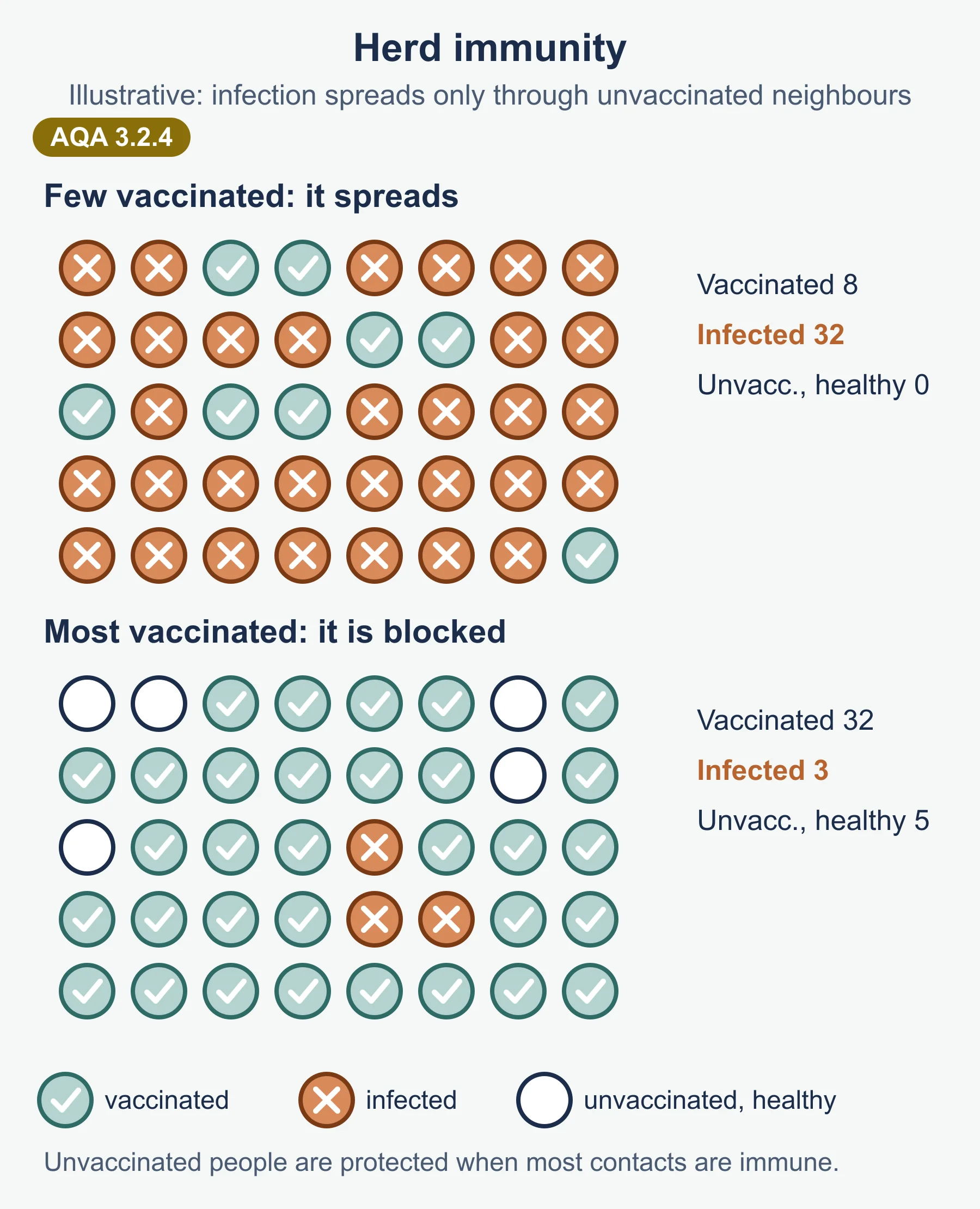
- For populations (herd immunity): when a high enough proportion of people are immune, the pathogen is unlikely to reach the few who are not, such as babies or people who cannot be vaccinated.
Active and passive immunity
| Active | Passive | |
|---|---|---|
| Antibodies | Made by the person’s own plasma cells | Come from outside the body |
| Memory cells | Yes | No |
| Speed | Takes time to develop | Immediate |
| Duration | Long-term | Short-term; the antibodies are broken down |
| Examples | Natural infection; vaccination | Antibodies crossing the placenta or in breast milk; injected antibodies (for example antitoxin) |
HIV and AIDS
Structure of HIV
| Part | Description |
|---|---|
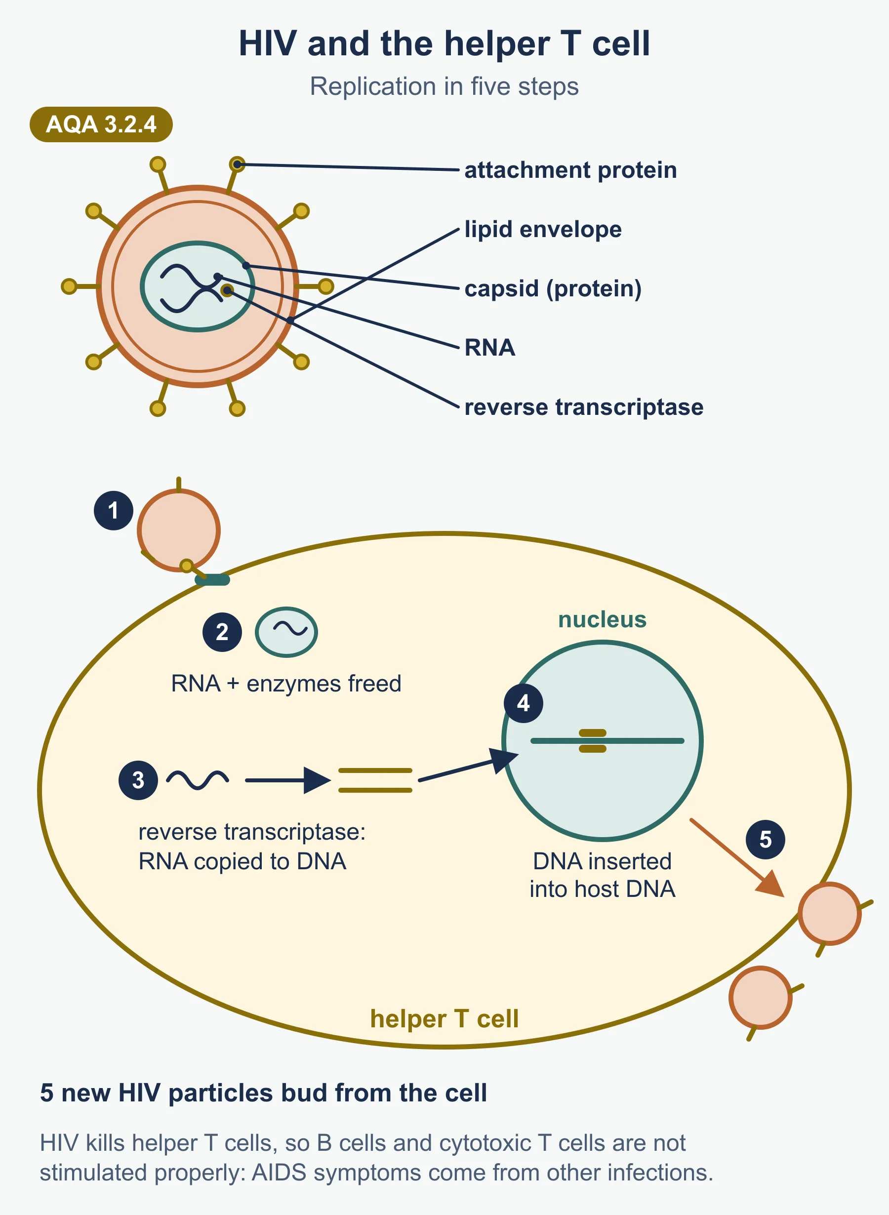
| Genetic material | RNA (two single strands) |
| Enzyme | Reverse transcriptase, packaged with the RNA |
| Capsid | Protein coat around the RNA and enzymes |
| Envelope | A lipid layer taken from the host cell membrane |
| Attachment proteins | In the envelope; bind to a receptor on helper T cells |
Replication in helper T cells
- Attachment proteins bind to a receptor protein on a TH cell.
- The capsid enters the cell and releases the RNA and reverse transcriptase.
- Reverse transcriptase makes DNA from the viral RNA.
- This DNA is inserted into the host cell’s DNA.
- The host cell transcribes it and makes viral proteins and RNA.
- New virus particles are assembled and bud from the cell, taking some of its membrane as their envelope.
How HIV causes the symptoms of AIDS
- HIV infects and destroys helper T cells, so their number falls.
- With few TH cells, B cells, cytotoxic T cells and phagocytes are not stimulated properly.
- The immune system cannot respond to other pathogens, so opportunistic infections (and some cancers) develop. These cause the symptoms of AIDS, not HIV itself.
Why antibiotics do not work against viruses
Antibiotics work by damaging processes that bacteria have, such as making murein cell walls or protein synthesis on bacterial ribosomes. Viruses have no cell wall, no ribosomes and no metabolism of their own, and they replicate inside host cells, so there is nothing for the antibiotic to act on.
Monoclonal antibodies
Details of how monoclonal antibodies are produced commercially are not required.
| Use | How it works |
|---|---|
| Targeting medication | A therapeutic drug is attached to an antibody complementary to an antigen found only on one cell type (for example a cancer cell). The drug is delivered to those cells, so fewer healthy cells are harmed and a lower dose may be needed |
| Medical diagnosis | Antibodies that bind a specific molecule show whether it is present, for example in pregnancy tests (detecting the hormone hCG in urine) or in tests for some cancers and infections |
The ELISA test
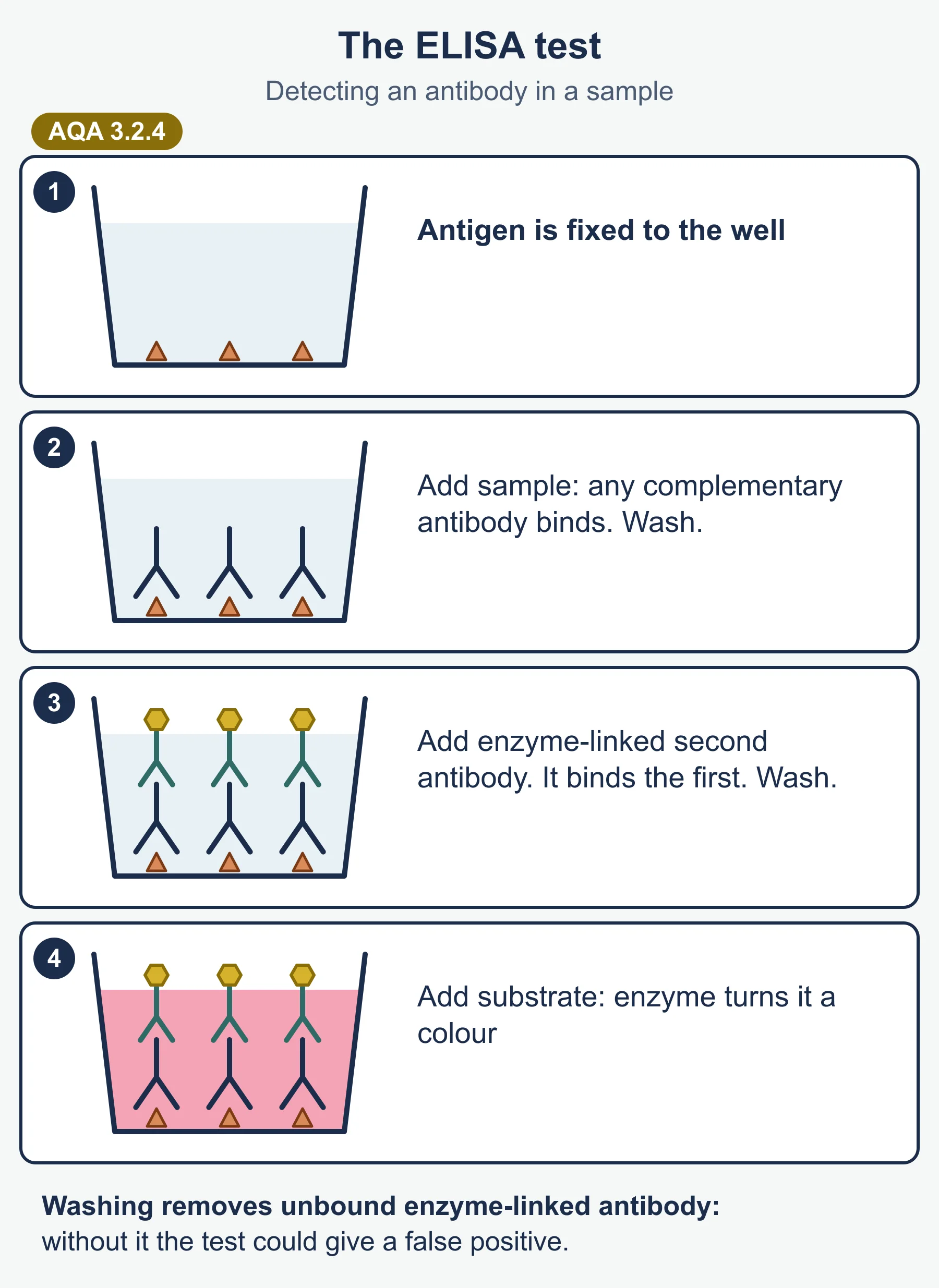
ELISA (enzyme-linked immunosorbent assay) uses antibodies linked to an enzyme to detect, and roughly measure, a particular antigen or antibody.
Indirect ELISA, for example testing blood for antibodies against HIV:
- HIV antigen is fixed to the bottom of a well.
- The patient’s blood plasma is added. If HIV antibodies are present, they bind the antigen.
- The well is washed to remove unbound antibodies.
- A second antibody, with an enzyme attached, is added; it binds to the patient’s antibodies.
- The well is washed again.
- A substrate is added. If the enzyme is present it produces a colour change, meaning a positive result. More colour means more antibody.
Direct ELISA detects an antigen: an antibody for the antigen is fixed to the well, the sample is added, then an enzyme-linked antibody that also binds the antigen, then the substrate.
Why wash? Unbound enzyme-linked antibody left in the well would change the colour and give a false positive. Run a control well without the patient sample to check this.
Ethical issues
Vaccines
- Testing may involve animals, and trials involve human volunteers who take some risk.
- Rare side effects; how to weigh small individual risks against large population benefits.
- Should vaccination be compulsory, or is it a personal choice? People who refuse lower herd immunity for others.
- Who gets a limited supply first, and who pays in poorer countries?
Monoclonal antibodies
- Their production has involved animals (mice are used to produce the antibody-making cells).
- Drug trials carry risk: in the 2006 TGN1412 trial, healthy volunteers given a new monoclonal antibody became seriously ill.
- Treatments can be very expensive, which raises questions about fair access.
When asked to evaluate data on vaccines or monoclonal antibodies, look at sample size, whether there was a control or placebo group, whether the trial was double blind, the length of follow-up, whether differences are significant, and who funded the study.
Must-know distinctions
- Antigen versus antibody. Antigens are the markers; antibodies are the proteins that bind them.
- Lysosome versus lysozyme. The lysosome is the organelle; lysozymes are the enzymes it releases.
- Clonal selection versus clonal expansion. Selection is choosing the cell with complementary receptors; it then divides by mitosis.
- Helper versus cytotoxic T cells. TH cells stimulate other cells; TC cells kill infected cells.
- Antibody and antigen are complementary, not “the same shape”.
- HIV versus AIDS. HIV is the virus; AIDS is the condition caused by the loss of TH cells.
Quick self-test
- Define antigen.
- List four things that cell-surface molecules allow the immune system to identify.
- Describe what happens after a pathogen has been engulfed by a phagocyte.
- Give three cell types that helper T cells stimulate.
- What are the variable regions of an antibody for?
- Explain why antibodies cause agglutination.
- Why does the secondary response produce antibodies faster than the primary response?
- Give one difference between active and passive immunity.
- What enzyme does HIV use to make DNA from its RNA?
- Why are antibiotics ineffective against HIV?
- Why is the well washed between stages in an ELISA test?
- Explain how herd immunity protects an unvaccinated baby.
Answers
- A molecule (usually a protein) that is recognised as foreign by the immune system and triggers an immune response.
- Pathogens, cells from other organisms of the same species, abnormal body cells and toxins.
- It is enclosed in a phagosome; a lysosome fuses with it and lysozymes hydrolyse the pathogen; the antigens are then presented on the phagocyte’s surface.
- Cytotoxic T cells, B cells and phagocytes.
- They form the antigen-binding sites, which are complementary to one specific antigen.
- Each antibody has two binding sites, so it can bind two bacteria, linking many into clumps.
- Memory cells with complementary receptors are already present, so they divide quickly into plasma cells.
- Active: the body makes its own antibodies and memory cells and protection is long-term. Passive: antibodies come from outside, there are no memory cells and protection is short-term.
- Reverse transcriptase.
- HIV has no cell wall, ribosomes or metabolism of its own and replicates inside host cells, so antibiotics have no target.
- To remove unbound antibodies, which would otherwise give a false positive result.
- Most people around the baby are immune, so the pathogen is unlikely to spread to the baby.
Where marks are usually lost
- Saying antibodies are “the same shape as” antigens rather than complementary.
- Writing that phagocytes “eat” bacteria, without naming the phagosome, lysosome and hydrolysis.
- Saying vaccines contain antibodies (that is passive immunity).
- Saying HIV directly kills people, rather than the opportunistic infections that follow loss of TH cells.
- Missing the washing step, or its reason, in ELISA answers.
- Forgetting that memory cells are the reason the secondary response is faster.
Frequently asked questions
Why are antibodies described as complementary to antigens? Each antibody’s binding site has a shape complementary to one specific antigen, so they fit together. Saying they are “the same shape” is a common error in AQA 7402 answers.
Why is the well washed in an ELISA test? Washing removes unbound antibodies. Any unbound enzyme-linked antibody left in the well would change colour and give a false positive.
How does HIV cause the symptoms of AIDS? HIV infects and destroys helper T cells, so B cells, cytotoxic T cells and phagocytes are not stimulated properly. The immune system cannot respond to other pathogens, and opportunistic infections cause the symptoms.
Why does antigen variability make people ill again? When a pathogen such as the influenza virus changes its surface antigens, memory cells from an earlier infection or vaccine no longer have complementary receptors, so the next infection triggers a slow primary response.
Do I need to know how monoclonal antibodies are produced for AQA? No. The specification states that details of the commercial production of monoclonal antibodies are not required.
Official syllabus
AQA AS and A-level Biology specification (7401 and 7402), for first teaching 2015, exams June 2016 onwards, Version 1.5, published by AQA. Section 3.2.4 Cell recognition and the immune system.
Get free revision emails (optional)
Occasional emails with practice questions, worked explanations and links to free resources for the qualification and subjects you choose. No spam, and you can unsubscribe from any email. The free tools on this site never need an email.
Related resources
-
Exam preparation
AQA A-Level Biology: Paper-by-Paper Exam Preparation
How AQA A-Level Biology 7402 Papers 1, 2 and 3 work, with strategy for the Paper 2 comprehension, the Paper 3 essay and practical skills, plus a checklist.
Biology · AQA · A Level
Read guide -
Study guides
AQA A-level Biology: Cell Recognition and the Immune System (7402)
Study guide to AQA A-level Biology 3.2.4: antigens, phagocytosis, T and B cells, antibodies, vaccines and herd immunity, HIV, monoclonal antibodies and ELISA.
Biology · AQA · A Level (AS content)
Read guide -
Practice questions
AQA A-level Biology: Cell Recognition and the Immune System (7402) – Practice Questions
Original questions with worked answers on AQA A-level Biology 3.2.4: antigens, phagocytosis, T and B cells, antibodies, vaccines, HIV, monoclonals and ELISA.
Biology · AQA · A Level (AS content)
Try questions
Related articles
-
study skills
How to revise for a science examination
Most science revision fails because it rereads notes instead of retrieving them. A practical method for revising physics, chemistry and biology in the weeks before a paper.
14 July 2026
-
teaching
A Level tuition in Dubai: Cambridge or Pearson Edexcel International A Level?
Schools in Dubai and Abu Dhabi teach either Cambridge International AS & A Level or Pearson Edexcel International A Level. How the two are structured, why it matters for tuition, and how online classes from Lahore work.
9 October 2026
Studying this with a teacher
Working through Biology A LEVEL (AS CONTENT)?
This page is free and stays free. If you would rather be taught it, Marlbridge runs Biology classes one-to-one and in small groups of up to 15, online in your own time zone. The first trial class is free. WhatsApp replies within an hour (8am–11pm Pakistan time, every day); email the same day.
AQA Biology teachers at Marlbridge


