
Revision Notes
OCR A Level Biology A: Cell division, diversity and organisation (H420) – Revision Notes
Condensed notes on the cell cycle, mitosis, meiosis, the PAG1 root tip squash, specialised cells and stem cells for OCR A Level Biology A H420, section 2.1.6.
- Subject
- Biology
- Level
- A LEVELS
- Topic
- Cell division, cell diversity and cellular organisation
- Author
- Marlbridge Academic Team
- Updated
- Reviewed by
- Hina Mogul (what this means)
Aligned to OCR A Level Biology (H420), Version 4.1 (April 2026), for first assessment in 2025. Official specification .
Syllabus page (what it covers and how it is assessed): OCR A Level Biology.
Syllabus points this page covers
H420
- 2.1.6 Cell division, cell diversity and cellular organisation
Found an error? Report a correction.
Need help with this topic? Request a free trial class for A Level Biology (H420).
These notes condense learning outcomes 2.1.6(a) to (m) of the OCR A Level Biology A specification (H420, Version 4.1, April 2026, first assessment 2025), including the PAG1 microscopy work on stained sections and squashes of plant tissue. The course sits on the OCR A Level Biology hub; tick off outcomes on the printable checklist. Microscope use, calibration and drawing rules are in the cell structure revision notes and the practical endorsement revision notes; DNA replication in S phase is in the nucleotides and DNA revision notes; and xylem and phloem return in the exchange and transport revision notes. The free diagnostics show gaps quickly.
Key definitions
| Term | Meaning |
|---|---|
| Cell cycle | The repeating sequence of growth, DNA replication and division that a dividing cell goes through |
| Chromatid | One of the two identical copies of a chromosome after replication, joined at the centromere |
| Centromere | The region where two sister chromatids are held together and where spindle fibres attach |
| Homologous chromosomes | A pair of chromosomes, one from each parent, of the same size and shape with the same genes at the same loci (possibly different alleles) |
| Diploid (2n) / haploid (n) | Two sets of chromosomes / one set of chromosomes |
| Differentiation | The process by which a cell becomes specialised for a particular function |
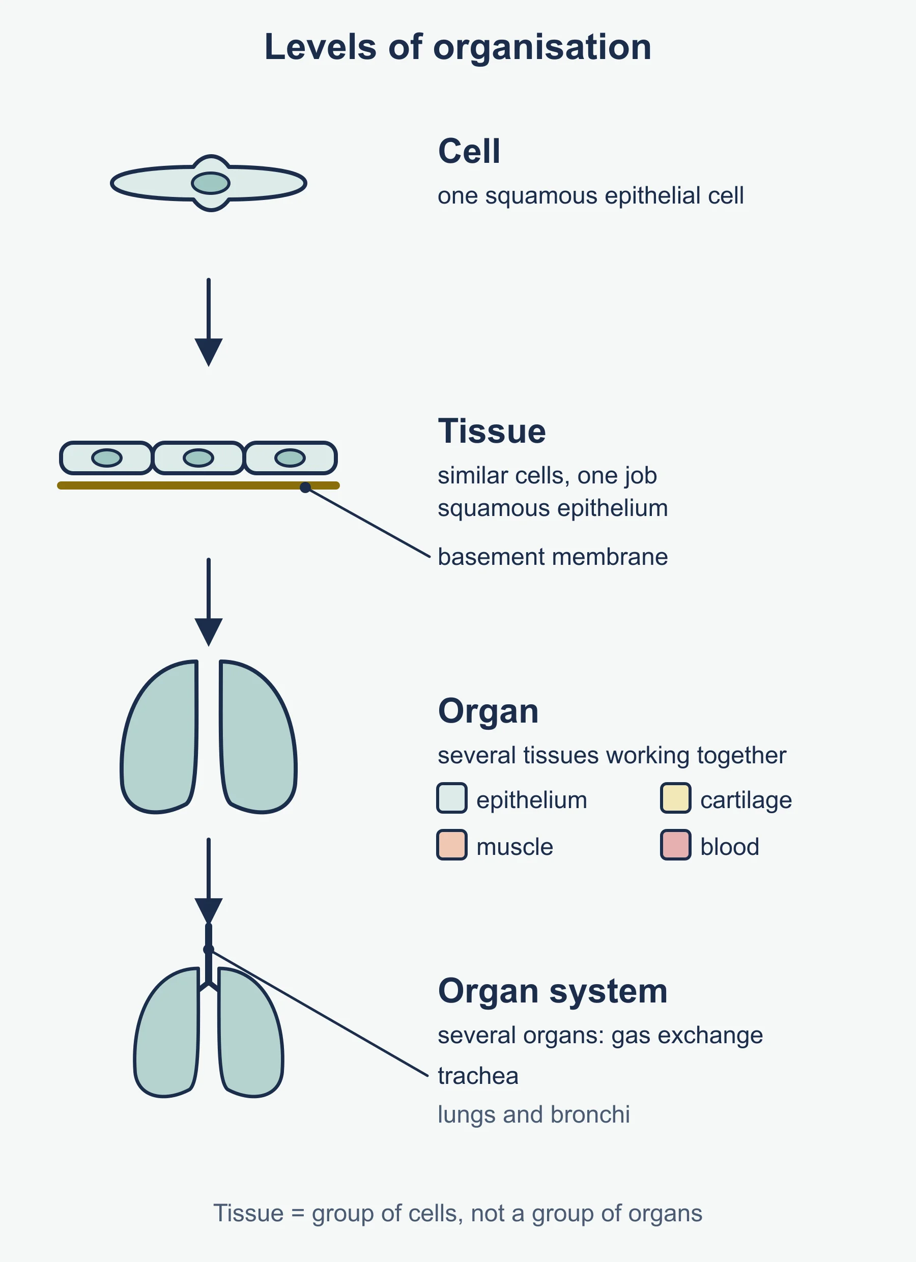
| Tissue | A group of similar cells, with any intercellular material, working together for a particular function |
| Organ | A group of different tissues working together to carry out a function |
| Organ system | A group of organs working together to carry out a major function |
| Stem cell | An undifferentiated cell that can divide repeatedly (self-renewal) and can differentiate into specialised cells |
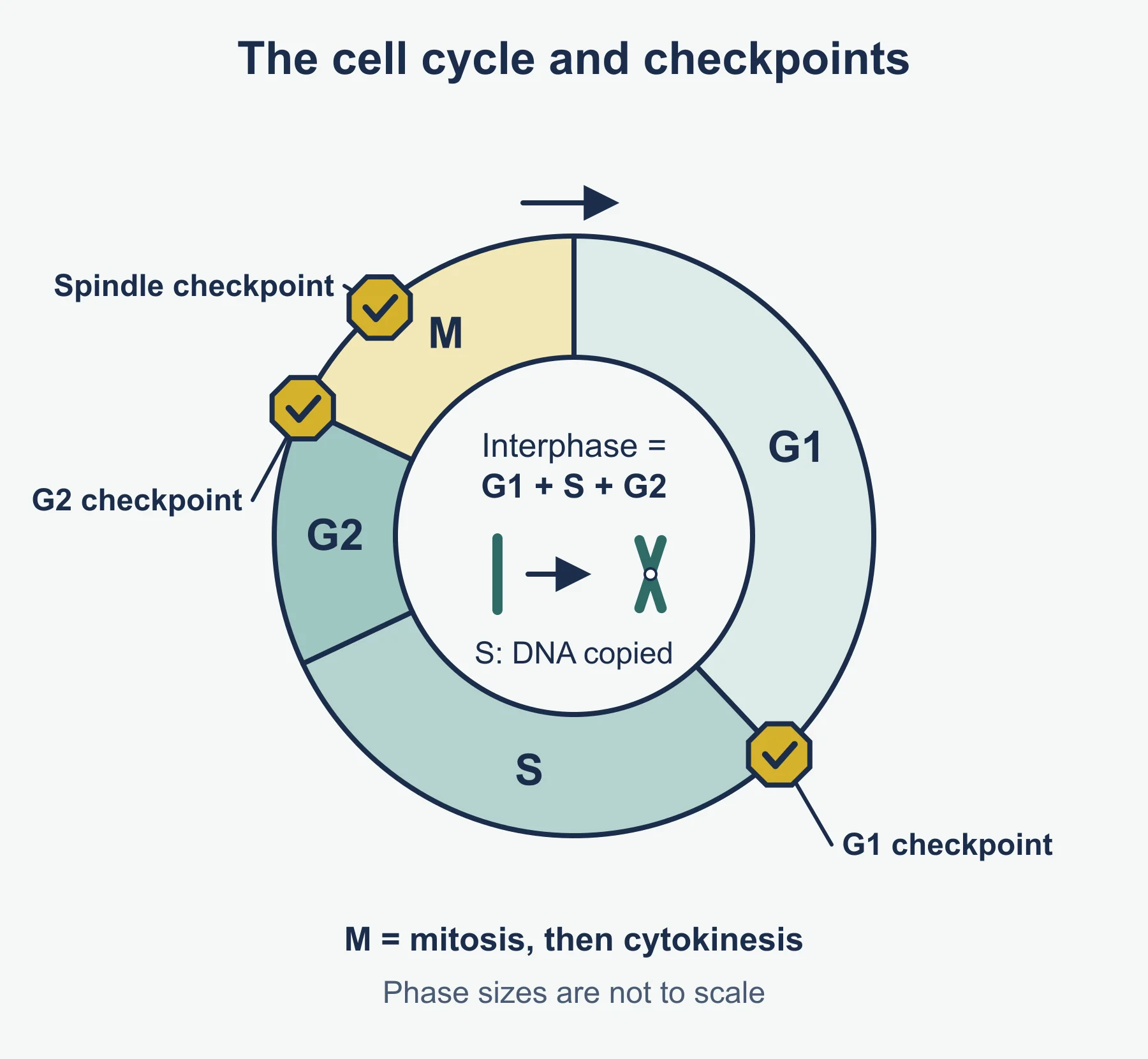
The cell cycle: 2.1.6(a)
| Stage | What happens |
|---|---|
| G1 (interphase) | Cell grows; proteins and organelles are made |
| S (interphase) | DNA is replicated, so each chromosome becomes two sister chromatids |
| G2 (interphase) | Further growth; energy stores built up; replicated DNA is checked for errors |
| Mitosis | The nucleus divides into two genetically identical nuclei |
| Cytokinesis | The cytoplasm divides, giving two genetically identical daughter cells |
- Interphase is the longest part of the cycle; the cell is not “resting” but very active.
- A cell can leave the cycle into G0, where it stops dividing, for example when it differentiates or becomes senescent.
- Mitosis gives genetically identical cells because DNA is copied exactly in S phase and the sister chromatids are shared equally.
Regulating the cell cycle: 2.1.6(b)
Checkpoints are control points where the cell is checked before it can move on.
| Checkpoint | When | Checks |
|---|---|---|
| G1 checkpoint | End of G1 | Cell size, nutrients, growth factors, DNA damage |
| G2 checkpoint | End of G2 | DNA fully replicated without damage; cell big enough to divide |
| Spindle assembly (metaphase) checkpoint | During mitosis | Every chromosome is attached to spindle fibres before anaphase starts |
- A cell that fails a checkpoint may pause for repair, enter G0 or undergo programmed cell death (apoptosis).
- If checkpoints fail, cells can divide uncontrollably, which can lead to a tumour.
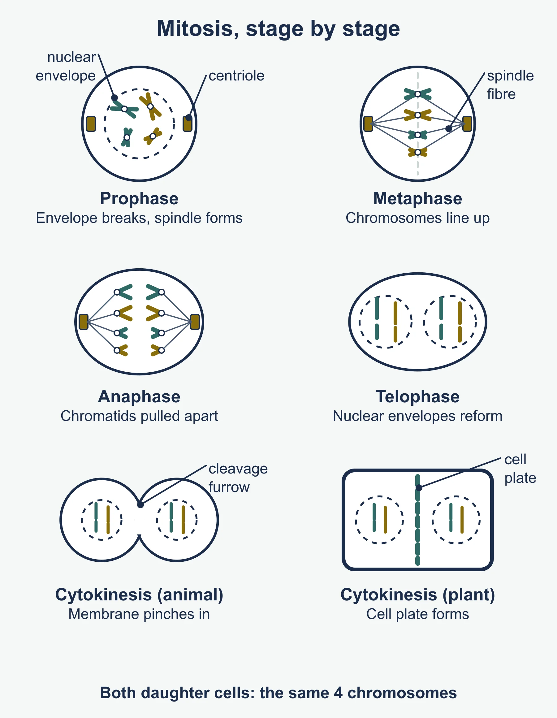
Mitosis: 2.1.6(c)
| Stage | Nuclear envelope | Chromosomes and chromatids | Centrioles and spindle |
|---|---|---|---|
| Prophase | Breaks down; nucleolus disappears | Chromatin condenses; chromosomes become visible as two sister chromatids joined at the centromere | Centrioles move to opposite poles (animal cells); spindle fibres form |
| Metaphase | Absent | Chromosomes line up at the equator, each attached by its centromere | Spindle fibres attached to centromeres |
| Anaphase | Absent | Centromeres divide; sister chromatids are pulled to opposite poles, centromere first (V shapes) | Spindle fibres shorten |
| Telophase | Reforms round each set of chromosomes; nucleoli reappear | Chromosomes uncoil back to chromatin | Spindle breaks down |
Cytokinesis
- Animal cells: the cell membrane is pulled inwards by the cytoskeleton to form a cleavage furrow, which pinches the cell in two.
- Plant cells: vesicles from the Golgi apparatus gather at the equator to form a cell plate, which becomes the new cell walls and membranes. Plant cells have no centrioles; their spindle still forms.
Memory order: PMAT, then cytokinesis.
Observing mitosis in plant tissue (PAG1): 2.1.6(d)
Root tip squash, in steps
- Cut the tips (a few millimetres) from growing roots of garlic or onion; the meristem there has many dividing cells.
- Warm in dilute hydrochloric acid for a few minutes; this breaks down the middle lamella so cells separate and stops division.
- Rinse in water, then place on a slide and add a stain that binds DNA (for example toluidine blue or acetic orcein) so chromosomes show up.
- Break up the tip with a mounted needle, add a coverslip and press down gently (with filter paper on top) to spread the cells into a single layer. Do not twist the coverslip.
- Find the cells at low power, then look at high power (×400).
- Prepared stained sections of root tips can be used in the same way.
- Labelled diagrams: clear continuous lines, no shading, label the stage and the chromosomes, spindle, cell wall and nuclear envelope where visible; include a title and magnification.
- Mitotic index = number of cells in mitosis ÷ total number of cells counted. A higher index means more active division.
- The proportion of cells in each stage reflects how long that stage lasts: most cells are in interphase.
Significance of mitosis: 2.1.6(e)
- Growth: more cells, all genetically identical, in plants, animals and fungi.
- Tissue repair and replacement: for example skin cells and the lining of the gut.
- Asexual reproduction: offspring are clones of the parent.
- Plants: runners, bulbs, tubers.
- Animals: budding in Hydra.
- Fungi: budding in yeast; spore production in many moulds.
Significance of meiosis: 2.1.6(f)
- Produces haploid cells (gametes in animals) from a diploid cell, so the diploid number is restored at fertilisation and the chromosome number stays constant between generations.
- Genetic variation by:
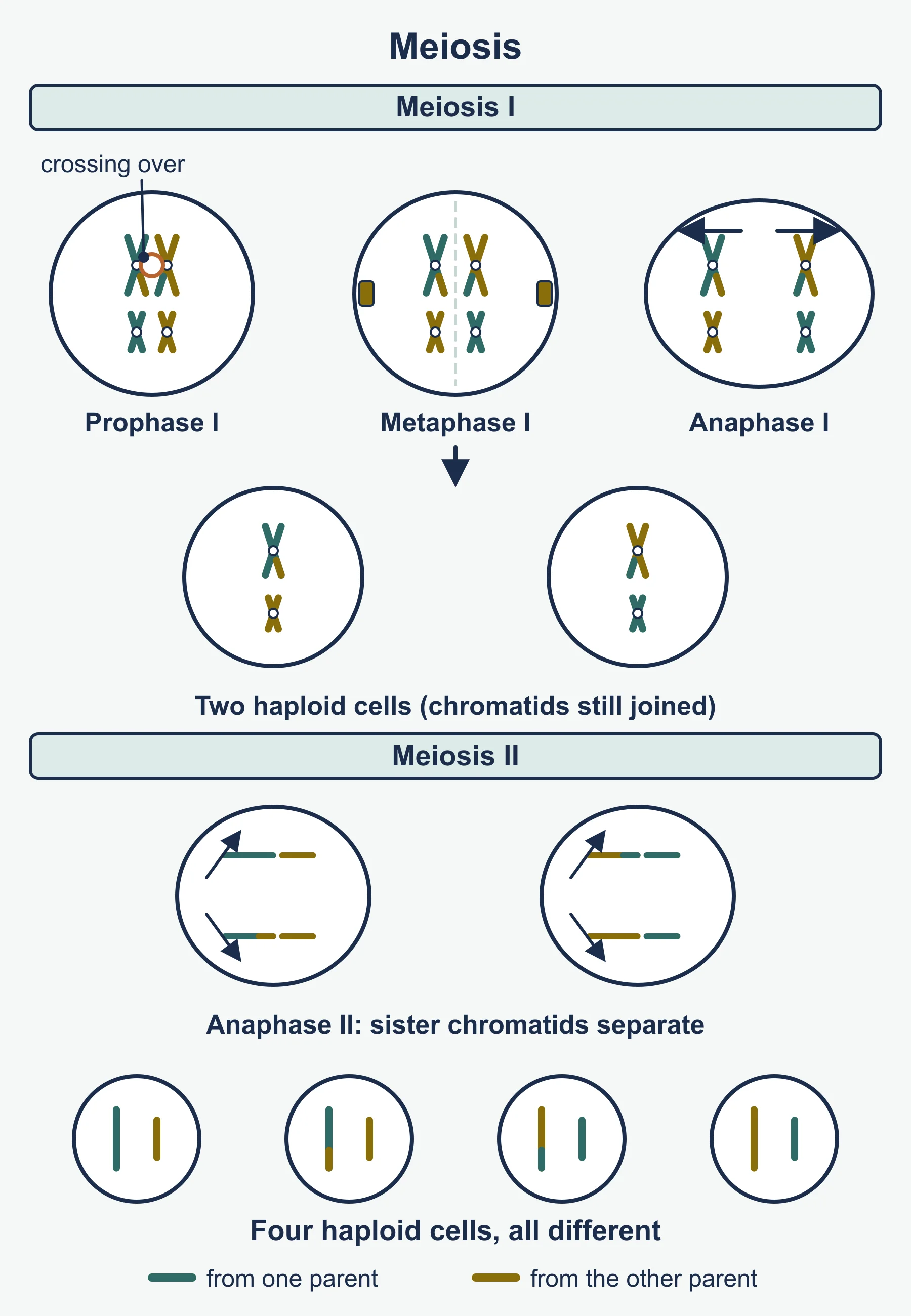
- Crossing over in prophase 1: non-sister chromatids of a homologous pair swap sections at chiasmata, making new combinations of alleles.
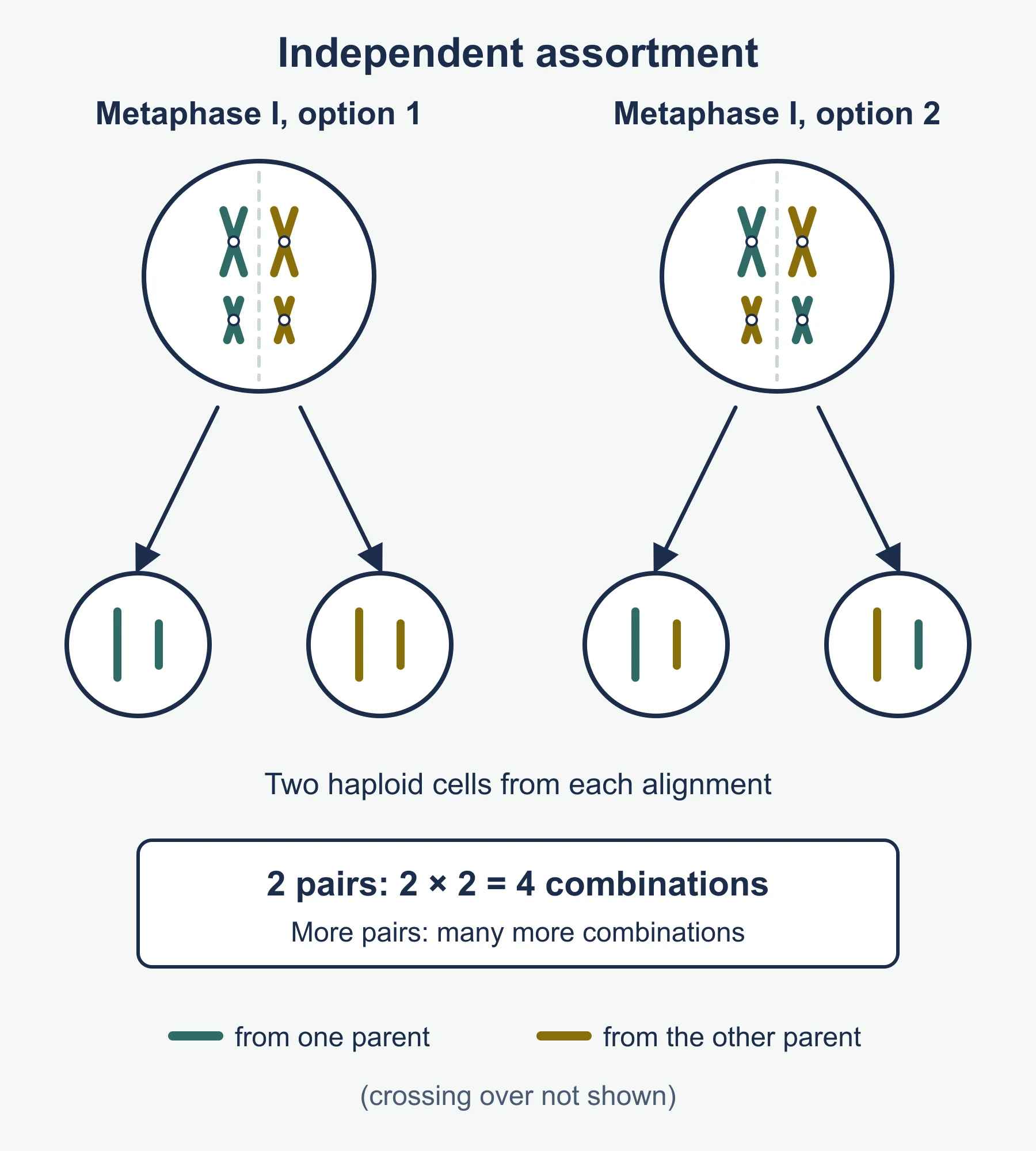
- Independent assortment in metaphase 1 (and of chromatids in metaphase 2): each homologous pair lines up independently, so maternal and paternal chromosomes are shuffled.
- Variation, together with random fertilisation, gives the differences that natural selection acts on.
Stages of meiosis: 2.1.6(g)
Meiosis has two divisions and gives four genetically different haploid cells. Interphase, with DNA replication, comes before it.
| Stage | Key events |
|---|---|
| Prophase 1 | Chromosomes condense; homologous chromosomes pair up (bivalents); crossing over at chiasmata; nuclear envelope breaks down; spindle forms |
| Metaphase 1 | Homologous pairs line up at the equator, randomly orientated (independent assortment) |
| Anaphase 1 | Homologous chromosomes are pulled to opposite poles; sister chromatids stay joined |
| Telophase 1 | Two haploid nuclei form; cytokinesis follows in most cells |
| Prophase 2 | Nuclear envelopes break down again; new spindles form; no further DNA replication |
| Metaphase 2 | Individual chromosomes line up at the equator; chromatids are assorted independently |
| Anaphase 2 | Centromeres divide; sister chromatids are pulled to opposite poles |
| Telophase 2 | Four haploid nuclei form; cytokinesis gives four cells |
No names of the sub-stages of prophase 1 are needed. Meiosis can also be observed in prepared slides of anthers (PAG1).
| Mitosis | Meiosis | |
|---|---|---|
| Divisions | One | Two |
| Daughter cells | Two, diploid | Four, haploid |
| Genetically | Identical to parent and each other | Different from parent and each other |
| Homologous pairs | Do not pair | Pair and cross over |
| Role | Growth, repair, asexual reproduction | Gamete production, variation |
Specialised cells: 2.1.6(h)
| Cell | Function | Adaptations |
|---|---|---|
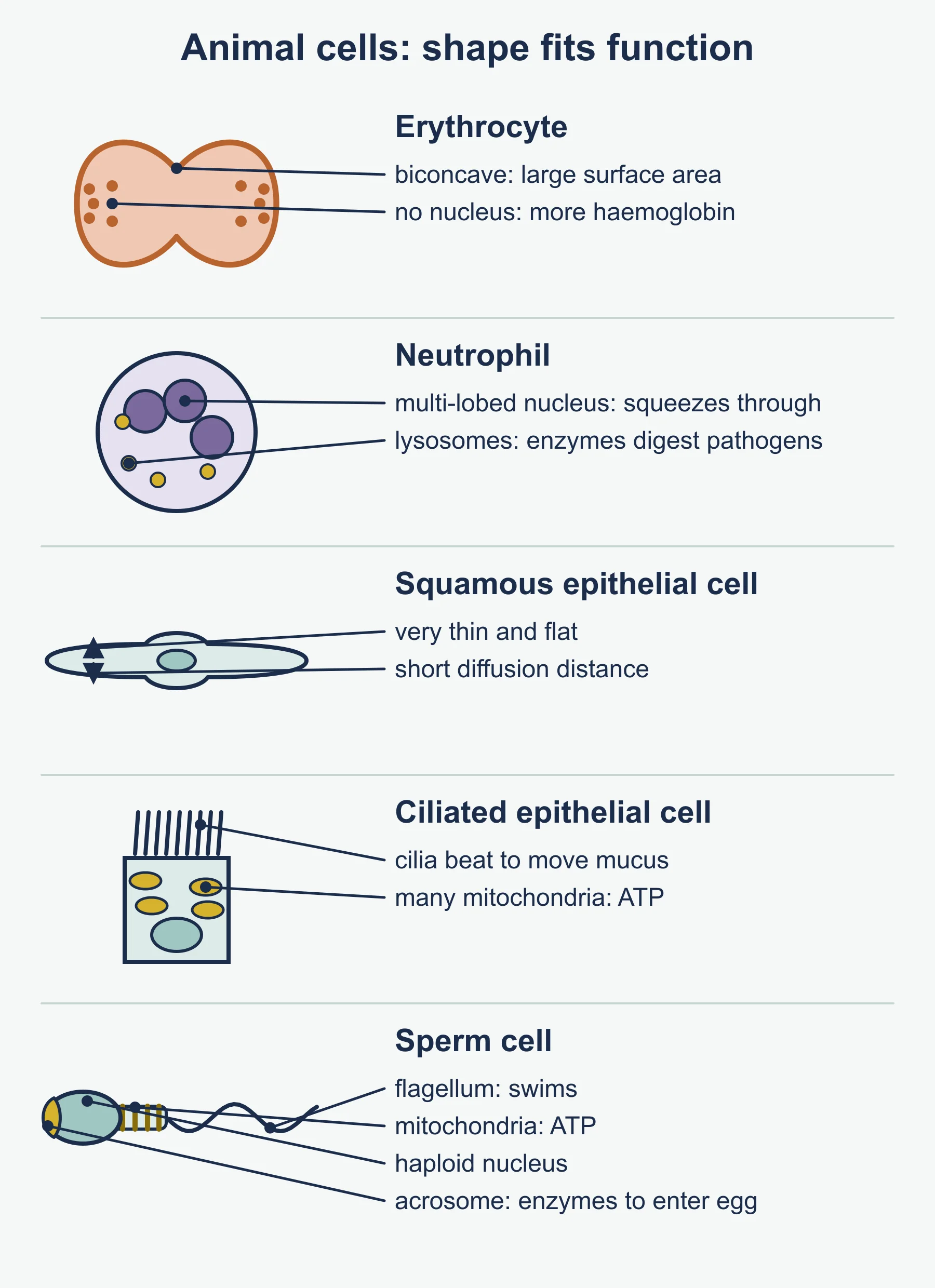
| Erythrocyte (red blood cell) | Carries oxygen | Biconcave disc: large surface area to volume ratio; no nucleus, so more room for haemoglobin; flexible to squeeze through capillaries |
| Neutrophil | Phagocytosis of pathogens | Multilobed nucleus, helping it squeeze through gaps in capillary walls; many lysosomes with digestive enzymes; flexible membrane for engulfing |
| Squamous epithelial cell | Lines surfaces such as alveoli | Very flat and thin: short diffusion distance |
| Ciliated epithelial cell | Lines the trachea and bronchi | Cilia beat to move mucus; many mitochondria supply ATP for this |
| Sperm cell | Delivers the male haploid nucleus | Flagellum for swimming; many mitochondria; acrosome with digestive enzymes to penetrate the egg; haploid nucleus |
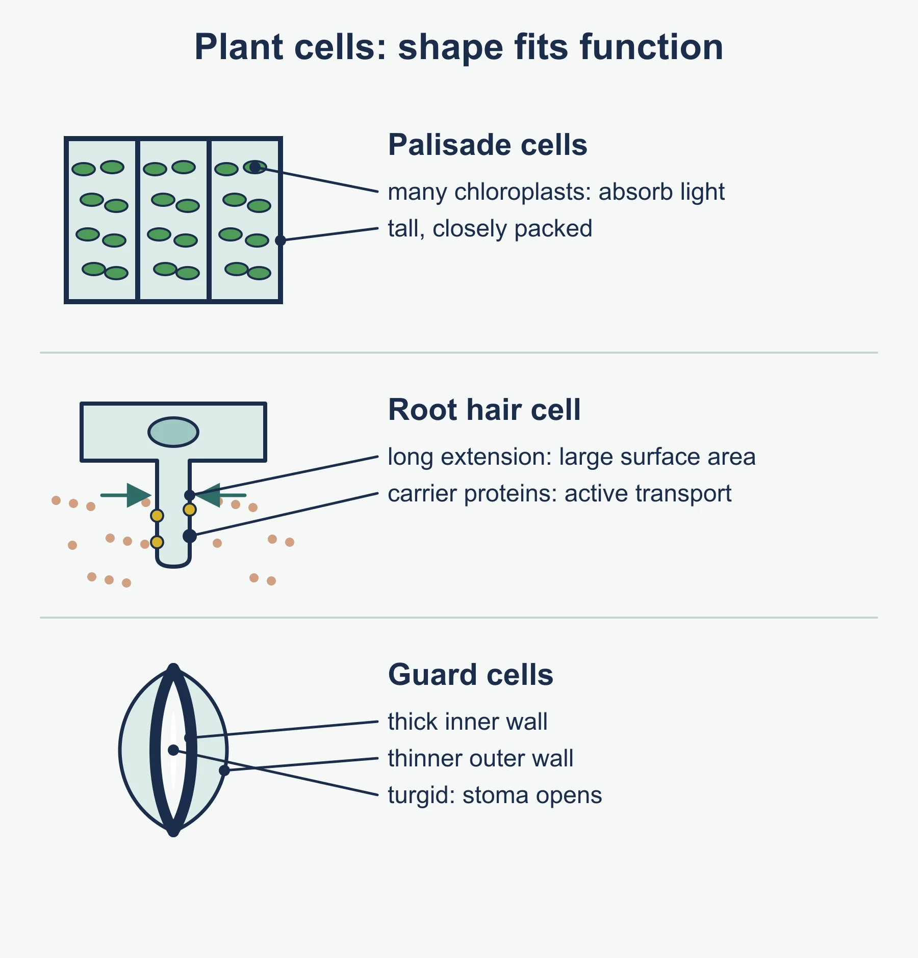
| Palisade cell | Photosynthesis | Many chloroplasts; tall, column shape packed near the upper leaf surface; large vacuole pushes chloroplasts to the edge |
| Root hair cell | Absorbs water and mineral ions | Long hair-like extension gives a large surface area; thin wall; many mitochondria for active transport of mineral ions |
| Guard cell | Opens and closes the stoma | Pair of cells; inner wall thicker than outer wall, so the cells bend apart when turgid; chloroplasts |
Tissues, organs and organ systems: 2.1.6(i)
| Tissue | Structure | Function |
|---|---|---|
| Squamous epithelium | One layer of flat cells on a basement membrane | Rapid diffusion, for example alveolar walls |
| Ciliated epithelium | Ciliated cells, with goblet cells secreting mucus | Moves mucus and trapped particles in the airways |
| Cartilage | Chondrocytes in a firm matrix containing collagen and elastin fibres | Support and flexibility, for example in the trachea, ear and ends of bones |
| Muscle | Cells or fibres that can contract (skeletal, smooth, cardiac) | Movement |
| Xylem | Dead, lignified vessels with no end walls, plus parenchyma and fibres | Transports water and mineral ions; support |
| Phloem | Sieve tube elements and companion cells | Transports assimilates such as sucrose |
- Organ examples: leaf, heart, lungs, stomach.
- Organ system examples: digestive, respiratory, circulatory (cardiovascular), and in plants the shoot and root systems.
- Cooperation between levels lets a multicellular organism carry out functions no single cell could.
Stem cells: 2.1.6(j)
- Stem cells are a renewing source of undifferentiated cells: they divide by mitosis, keeping some cells as stem cells while others differentiate.
- Differentiation happens because different genes are switched on or off, so different proteins are made.
| Potency | Can become | Example |
|---|---|---|
| Totipotent | Any cell type, including placental cells | Zygote and early embryo cells |
| Pluripotent | Any body cell type (not placenta) | Embryonic stem cells |
| Multipotent | A range of cell types within one family | Bone marrow (haematopoietic) stem cells |
| Unipotent | One cell type | Stem cells in the testes that produce only sperm |
Sources: embryos, adult tissues (bone marrow), umbilical cord blood, and induced pluripotent stem (iPS) cells made by reprogramming adult cells. In plants, stem cells are in the meristems: at root and shoot tips (apical meristems) and in the vascular cambium.
Erythrocytes and neutrophils from bone marrow: 2.1.6(k)
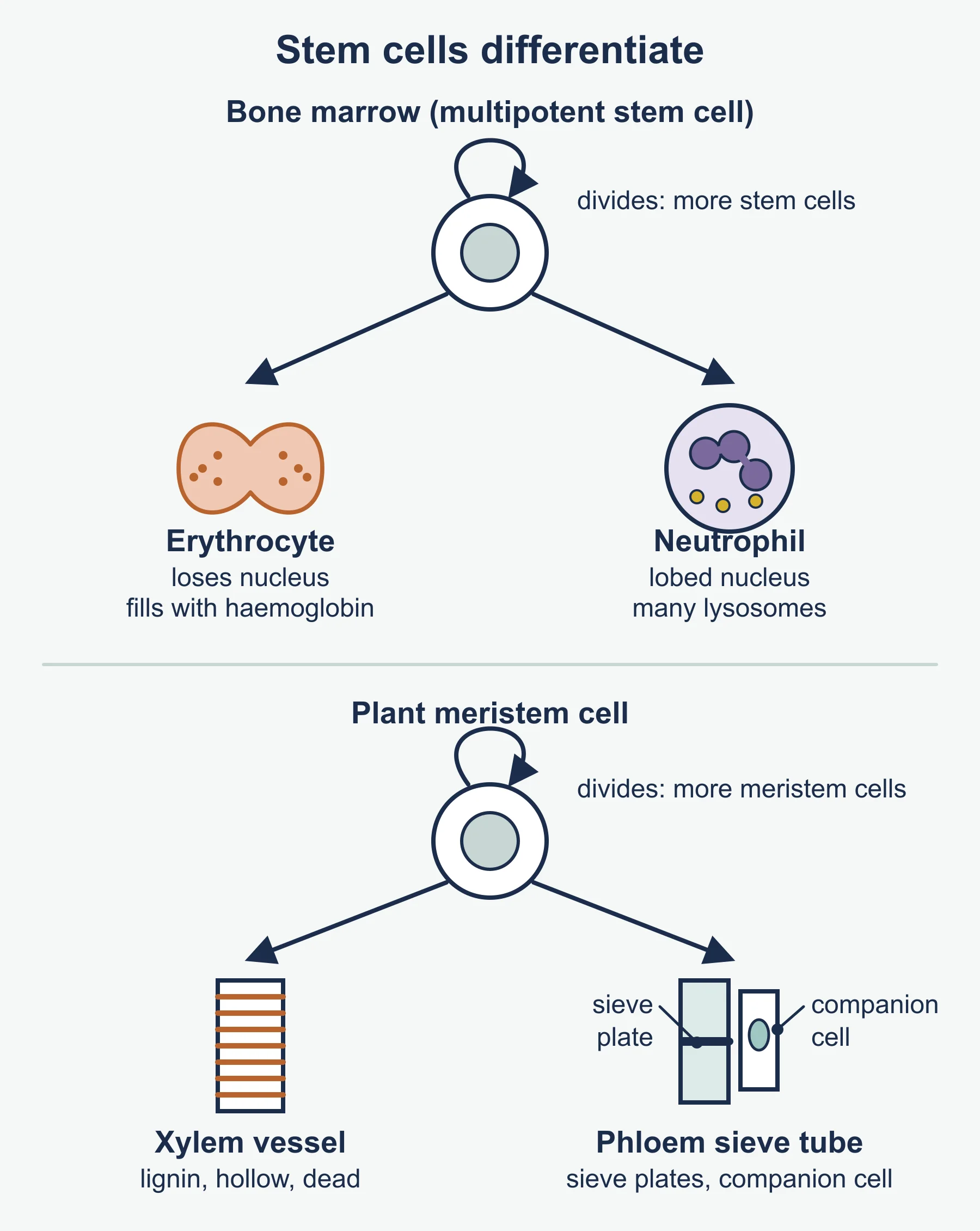
- Both come from the same multipotent stem cells in bone marrow, which produce all the blood cell types.
- Erythrocytes: the developing cells fill with haemoglobin and lose their nucleus, mitochondria, Golgi apparatus and ER, making room for more haemoglobin. They live about 120 days, so they are constantly replaced.
- Neutrophils: develop a multilobed nucleus and many lysosomes, and keep their organelles. They are short-lived and made in large numbers.
- Both differentiate along different routes because different genes are expressed.
Xylem and phloem from meristems: 2.1.6(l)
- In the vascular cambium, dividing meristem cells produce xylem cells on the inside and phloem cells on the outside.
- Xylem vessel elements: cells elongate; lignin is laid down in their walls (spiral, ring or more continuous patterns); the cells die and the contents and end walls break down, forming continuous hollow tubes. Pits remain where lignin is absent.
- Phloem sieve tube elements: lose their nucleus and most organelles; the end walls become sieve plates with pores. Each is linked by plasmodesmata to a companion cell, which keeps its nucleus and many mitochondria and supports the sieve tube element.
Uses of stem cells: 2.1.6(m)
- Repair of damaged tissues: for example bone marrow transplants to replace blood cells, and research into repairing damaged heart muscle, skin or cartilage.
- Treating neurological conditions: replacing lost or damaged nerve cells (no specific conditions need to be recalled).
- Developmental biology research: studying how cells differentiate and how organisms develop, and testing new drugs on tissues grown from stem cells.
| Issue | Points to weigh |
|---|---|
| Embryonic stem cells | Pluripotent and versatile, but collecting them destroys the embryo, which many people object to |
| Adult and iPS cells | Avoid destroying embryos; adult cells are only multipotent; iPS cells are still being refined |
| Safety | Risk of rejection, infection, or uncontrolled division leading to tumours |
| Regulation | Research is licensed and controlled by law in many countries |
Worked reminders
Mitotic index. 36 of 240 cells counted are in mitosis: 36 ÷ 240 = 0.15 (15 %).
Time in a stage. The cycle lasts 20 hours and 5 % of cells are in metaphase: 0.05 × 20 = 1 hour, about 60 minutes.
Chromosome numbers. A human cell (2n = 46): after mitosis each daughter has 46; after meiosis each gamete has 23.
Independent assortment. With n pairs of homologous chromosomes, the number of combinations is 2ⁿ. For n = 3: 2³ = 8. For humans (n = 23): 2²³ = 8 388 608, before crossing over is counted.
DNA content. If a G1 nucleus holds x units of DNA, a G2 nucleus holds 2x; each daughter after mitosis holds x; each cell after meiosis holds 0.5x.
Writing good answers
- Name the structures the specification lists for each stage: nuclear envelope, chromosomes, chromatids, centromere, centrioles, spindle fibres, cell membrane.
- Separate homologous chromosomes from chromatids: in anaphase 1 homologues separate; in anaphase 2 (and in mitosis) chromatids separate.
- Link adaptation to function in one sentence: “No nucleus, so more space for haemoglobin, so more oxygen carried”.
- Explain differentiation through genes: different genes are expressed, so different proteins are made.
- Balance a stem cell question: give a benefit and an ethical or safety issue.
Must-know distinctions
- Chromosome versus chromatid versus homologous chromosome.
- Mitosis (one division, identical diploid cells) versus meiosis (two divisions, varied haploid cells).
- Crossing over (prophase 1) versus independent assortment (metaphase 1 and 2).
- Animal cytokinesis (cleavage furrow) versus plant cytokinesis (cell plate).
- Tissue versus organ versus organ system.
- Totipotent versus pluripotent versus multipotent.
- Xylem (dead, lignified, water) versus phloem (living sieve tubes, assimilates).
Quick self-test
- What happens in S phase?
- What does the spindle assembly checkpoint check?
- Describe the chromosomes at metaphase of mitosis.
- What happens to the centromeres in anaphase of mitosis?
- Why is root tip tissue treated with hydrochloric acid before squashing?
- Give three roles of mitosis in life cycles.
- Name the two processes in meiosis that produce genetic variation, and when each happens.
- What separates in anaphase 1?
- Give two adaptations of an erythrocyte.
- Why does a neutrophil have a multilobed nucleus?
- Name two tissues found in the H420 list that are plant tissues.
- Define a stem cell.
- Where are the stem cells that produce erythrocytes and neutrophils?
- How does a xylem vessel element form from a meristem cell?
- A count gives 18 cells in mitosis out of 150. Calculate the mitotic index.
Answers
- DNA is replicated, so each chromosome becomes two sister chromatids.
- That every chromosome is attached to spindle fibres before anaphase begins.
- They line up at the equator, each made of two sister chromatids, attached to spindle fibres by the centromere.
- They divide, so the sister chromatids separate and are pulled to opposite poles.
- To break down the middle lamella so the cells separate and spread into a single layer (it also stops division).
- Growth, tissue repair, asexual reproduction.
- Crossing over in prophase 1; independent assortment in metaphase 1 (and metaphase 2).
- The homologous chromosomes of each pair.
- Any two: biconcave shape, no nucleus, flexible, contains haemoglobin.
- So it can squeeze through gaps in capillary walls to reach infected tissue.
- Xylem and phloem.
- An undifferentiated cell that can divide repeatedly and differentiate into specialised cell types.
- In the bone marrow.
- It elongates, its walls are lignified, the cell dies and its contents and end walls break down to form a hollow tube.
- 18 ÷ 150 = 0.12.
Where marks are usually lost
- Calling interphase a “resting stage”, or saying DNA is replicated during prophase.
- Saying homologous chromosomes are identical; they carry the same genes but may carry different alleles.
- Writing that chromatids separate in anaphase 1.
- Giving “spindle fibres form” without saying they attach to the centromeres.
- Saying meiosis halves the chromosome number “to make gametes” without saying why (to keep the number constant at fertilisation).
- Twisting the coverslip in a squash, so chromosomes are smeared, or drawing cells with shading.
- Calling a tissue an organ (for example “xylem is an organ”).
- Saying stem cells are found only in embryos, or forgetting that plant stem cells are in meristems.
Frequently asked questions
What happens in the S phase of the cell cycle? In the S phase of interphase, DNA is replicated, so each chromosome becomes two sister chromatids. Interphase is the longest part of the cycle and the cell is very active, not resting.
How do you calculate the mitotic index? Divide the number of cells in mitosis by the total number of cells counted. For example, 36 cells in mitosis out of 240 gives a mitotic index of 0.15, or 15%.
How many chromosome combinations can independent assortment produce? With n pairs of homologous chromosomes, independent assortment gives 2ⁿ combinations. For humans, with 23 pairs, that is 2²³, before crossing over is counted.
What are the sources of stem cells? Stem cells come from embryos, adult tissues such as bone marrow, umbilical cord blood and induced pluripotent stem (iPS) cells made by reprogramming adult cells. In plants, stem cells are found in the meristems.
Official syllabus
OCR Level 3 Advanced GCE in Biology A (H420) specification, Version 4.1 (April 2026), first assessment 2025, published by OCR. Module 2.1.6 Cell division, cell diversity and cellular organisation, statements (a) to (m), with PAG1 (microscopy).
Get free revision emails (optional)
Occasional emails with practice questions, worked explanations and links to free resources for the qualification and subjects you choose. No spam, and you can unsubscribe from any email. The free tools on this site never need an email.
Related resources
-
Study guides
OCR A Level Biology A: Cell division, diversity and organisation (H420)
OCR A Level Biology A (H420) 2.1.6 study guide: cell cycle and checkpoints, mitosis, meiosis, specialised cells, tissues and organs, stem cells and their uses.
Biology · OCR · A Level
Read guide -
Practice questions
OCR A Level Biology A: Cell division, diversity and organisation (H420) – Practice Questions
Original OCR A Level Biology A (H420) 2.1.6 questions with worked answers on the cell cycle, mitosis, meiosis, specialised cells, tissues and stem cells.
Biology · OCR · A Level
Try questions -
Study guides
OCR A Level Biology A: Cell structure, microscopy, prokaryotes and eukaryotes (H420)
Study guide: microscopy, magnification, organelles, cytoskeleton and prokaryotes versus eukaryotes for OCR A Level Biology A (H420) with worked examples.
Biology · OCR · A Level
Read guide
Related articles
-
study skills
How to revise for a science examination
Most science revision fails because it rereads notes instead of retrieving them. A practical method for revising physics, chemistry and biology in the weeks before a paper.
14 July 2026
-
teaching
A Level tuition in Dubai: Cambridge or Pearson Edexcel International A Level?
Schools in Dubai and Abu Dhabi teach either Cambridge International AS & A Level or Pearson Edexcel International A Level. How the two are structured, why it matters for tuition, and how online classes from Lahore work.
9 October 2026
Studying this with a teacher
Working through Biology A LEVELS?
This page is free and stays free. If you would rather be taught it, Marlbridge runs Biology classes one-to-one, online in your own time zone. One-to-one only for this course: Rs 6,000 per class in Pakistan, with other currencies on the fees page. The first trial class is free. WhatsApp replies within an hour (8am–11pm Pakistan time, every day); email the same day.
OCR Biology teachers at Marlbridge


